
- Title
-
The interaction of epithelial Ihha and mesenchymal Fgf10 in zebrafish esophageal and swimbladder development
- Authors
- Korzh, S., Winata, C.L., Zheng, W., Yang, S., Yin, A., Ingham, P., Korzh, V., and Gong, Z.
- Source
- Full text @ Dev. Biol.

Endoderm-specific expression of ihha in early zebrafish development up to 96 hpf. A, A1, Expression of ihha is initiated in the endoderm at the level of 2–3 somites at 24 hpf. A1, Dorsal view. B, Expression of ihha increased at 36 hpf; B¹, lateral view shows a prominent domain of ihha expression in the foregut. C, D, At 50 hpf ihha expression is strong in the esophagus, swimbladder, liver, pneumatic (arrowhead) and extrahepatic ducts (arrows), lateral view. Caudal to the extrahepatic duct expression is in the region giving rise to the primitive intestine; E, E¹, by 74 hpf, ihha expression continues to be strong along the gastrointestinal tract in the esophagus and intestine, and in the pneumatic duct and swimbladder, but declines in the liver at 96 hpf, F, A–F whole mount. G–I, Cross-sections at 74 hpf show expression of ihha in the epithelium of the esophagus, pneumatic duct, swimbladder and intestine. The level of sections in G–I is shown by white lines in E. J, Schematic representation of ihha expression in the esophagus, gut, pneumatic duct and swimbladder. Unless otherwise stated anterior is to the left for all whole mounts. Abbreviations: e, endoderm; es, esophagus; ehd, extrahepatic duct; g, gut; l, liver; pd, pneumatic duct; sb, swimbladder; s, somite; y, yolk; *, midline. Scale bars: 250 μm. |
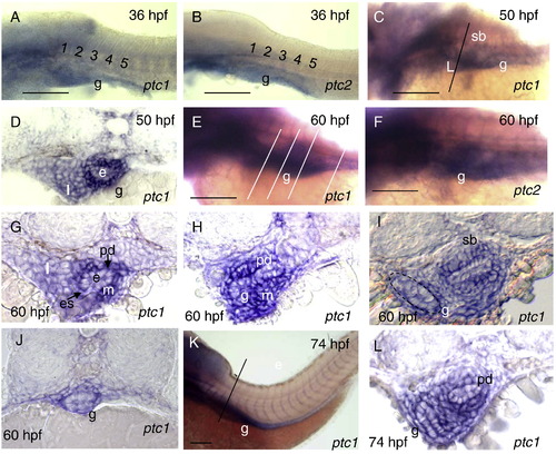
Expression pattern of ptc1 and ptc2 during development of the gut, pneumatic duct and swimbladder. A, B, Expression of ptc1 and ptc2 is detected by WISH at 36 hpf, C, ptc1 expression increased along the foregut and swimbladder bud at 50 hpf. A–C, Whole mount, lateral view. D, Cross-section of embryos in C revealed strong expression in the esophagus mesenchyme. E, F Expression of ptc1 and ptc2 is strong at 60 hpf. G–I, Cross-sections showed ptc1 expression domain in the mesenchyme surrounding epithelium of the esophagus, pneumatic duct, swimbladder and intestine. Note strong expression in the esophagus and along pneumatic duct. J, Cross section shows ptc1 expression in the posterior gut mesenchyme at 60 hpf. K, Expression of ptc1 at 74 hpf. L, Cross-section in K revealed strong expression of ptc1 in the mesenchyme surrounding the intestinal bulb, pneumatic duct and swimbladder. Abbreviations: e, epithelium; es, esophagus; g, gut, n, notochord; m, mesenchyme; pd, pneumatic duct; l, liver; sb, swimbladder. Scale bars: 250 μm. EXPRESSION / LABELING:
|
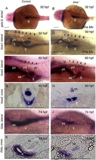
Morphogenesis of the gastrointestinal epithelium in ihha-/- mutant. A, B, Expression of endodermal marker gata6 at 32 hpf. C, D, Expression of foxa1 along the pharynx, esophagus, intestina, swimbladder and liver epithelium at 50 hpf. Note a reduced endoderm in the absence of Ihha signaling. E, F, At 60 hpf foxa2 is expressed in the pharynx, esophagus, swimbladder, pneumatic duct, intestine, and floor plate. foxa2 expression is not affected along the floor plate in ihha-/- mutant, but reduced in endodermal domains, A–F, whole mount. G, H, Cross sections of foxa2 expression revealed reduced epithelium of the intestine and swimbladder in ihha-/- compared to control embryos at 60 hpf. I, J, Expression of the enterocyte marker ifabp along gastrointestinal tract is altered in ihha-/- at 74 hpf. K, L, In control larvae strong expression of ifabp is in the apical aspect of the gastrointestinal epithelium demonstrating epithelial differentiation, which is less obvious in ihha-/-. Abbreviations: e, epithelium; L, liver; ph, pharynx; pd, pneumatic duct; es, esophagus; g, gut; ph, pharynx; s, somites; sb, swimbladder; n, notochord. Numbers represent anterior somites. Scale bars: 250 μm. EXPRESSION / LABELING:
PHENOTYPE:
|
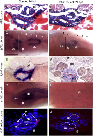
Morphogenesis of gastrointestinal tract mesenchyme in ihha-/-. A, B, cross sections with H & E staining show gastrointestinal tract of control and mutant larvae at the level of anterior swimbladder. Red lines show the size of epithelial and mesenchymal layers. Dotted red lines show the size of the lumen with epithelial and mesenchymal (arrow) layers in control and mutant larvae at 74 hpf. Note slightly reduced epithelial and mesenchymal layers of the gut and gut lumen in the mutant. C–F, Expression of fgf10a is prominent in anterior gut of control and reduced in mutant at 74 hpf. E, F, Cross sections show strong expression of fgf10a in the mesenchyme of esophagus in controls and significantly reduced in the corresponding region of ihha-/-. G, H, Expression of acta2 in the gastrointestinal tract mesenchyme. In ihha-/- mutant this expression is missing. I, J, Cross-section of gastrointestinal tract at the level of the anterior part of swimbladder in controls and ihha-/- reveals cell bodies of HuC/D-positive enteric neurons alongside of esophagus and intestine in controls and their absence in ihha-/- mutant. Dotted white lines separate epithelium from mesenchyme. Abbreviations: e, epithelium; es, esophagus; en, enteric neurons; m, mesenchyme; l, liver; sb, swimbladder; n, notochord. Numbers represent somites. Scale bars: 250 μm. EXPRESSION / LABELING:
PHENOTYPE:
|

Ihha is required for cell proliferation in the gastrointestinal tract. A–F, Proliferation assay using PCNA antibody. Cell nuclei are visualized with DAPI staining. A–F, Cross-section of gastrointestinal tract at the level of anterior part of swimbladder in A–C, controls and D–F, ihha-/- reveals decreased cells proliferation in both epithelial and mesenchymal layers of intestinal bulb in ihha-/- mutant. Dotted white lines demarcate epithelium and mesenchyme of intestinal bulb in control and ihha-/- mutant. G, Evaluation of epithelial and mesenchymal cell proliferation in the esophagus/intestinal bulb, mid-gut, and caudal gastrointestinal segments of control and ihha-/-. Epithelial cell proliferation in the absence of Ihha signaling is reduced significantly in the esophagus/intestinal bulb segment of ihha-/-. Most significant decrease in cell proliferation was observed in the mesenchyme of esophagus/intestinal bulb and mid-gut segments in ihha-/- comparing to controls. Solid red bars represent epithelium of esophagus/intestinal bulb, mid-gut and caudal gastrointestinal segments of control, while empty red boxes correspond to ihha-/-. Solid blue bars represent mesenchyme of esophagus/intestinal bulb, mid-gut and caudal gut of control, while others correspond to ihha-/-. There are no significant differences in cell proliferation in epithelium and mesenchyme of posterior gut in the control and ihha-/-. Abbreviations: e, epithelium; g, gut; sb, swimbladder. Scale bars: 250 μm. EXPRESSION / LABELING:
PHENOTYPE:
|
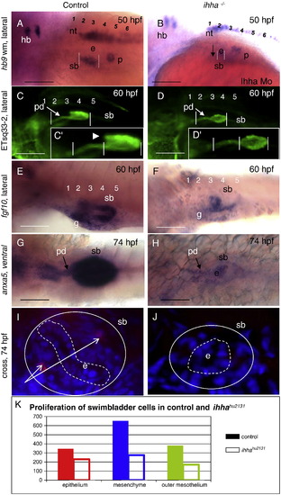
Defects of swimbladder development in the absence of Ihha. A, B, In control embryos expression of hb9, a marker for swimbladder epithelium, is initiated at 36 hpf and is expanded at 50 hpf. In Ihha morphant expression of hb9 in the swimbladder is delayed until 40–50 hpf, but expression of this marker elsewhere (the hindbrain, neural tube and endocrine pancreas) is unaffected. Note the same size of endocrine pancreas in control and Ihha morphant. C–D′, Swimbladder epithelia and pneumatic duct extensions (arrow) are significantly reduced in ihha-/-. Note that the anterior chamber (arrowhead) is absent in ihha-/- at 60 hpf. E, F, In control fgf10a-positive mesenchyme surrounded the epithelial bud starting from 48 hpf and increased at 60 hpf, in ihha-/- specification of swimbladder mesenchyme is initiated at around 60 hpf. G, H, Mesothelial domain of anxa5 in mutant larvae is almost absent, this staining also shows reduced swimbladder pneumatic duct and epithelium in mutant compared to control at 74 hpf. Anterior is to the left. I, J, Cross-section of swimbladder. HuC/D positive differentiated enteric neuron cell bodies are present in the mesenchymal layer of control larvae and absent along swimbladder mesenchyme in ihha-/-. Dotted white lines demarcate swimbladder epithelium. Solid white lines show the swimbladder size in control and ihha-/- larvae. K, assessment of cell numbers in swimbladder epithelium, mesenchyme and outer mesothelium of control and mutant. Solid colored bars correspond to control, while others correspond to ihha-/-. Abbreviations: hb, hindbrain; e, swimbladder epithelium; en, enteric neurons; p, endocrine pancreas; pd, pneumatic duct; n, notochord; nt, neural tube, s, somites; sb, swimbladder; g, gut. Numbers represent anterior somites. EXPRESSION / LABELING:
PHENOTYPE:
|
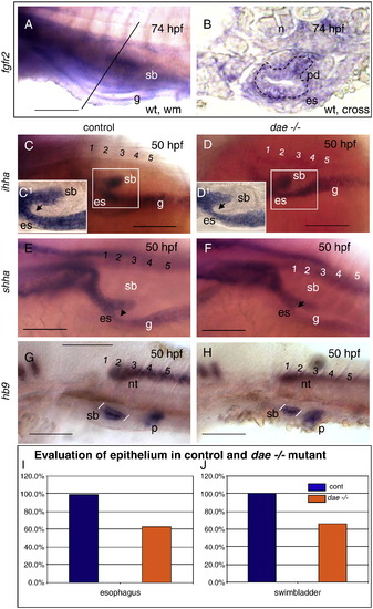
Endodermal phenotype of daetbvbo mutant. A, B, Expression of fgfr2 in the gut and swimbladder at 74 hpf. A, Whole mount. B, Cross section, expression of fgfr2 is in the epithelium of esophagus and pneumatic duct. C, D, Whole mount, expression of ihha in dae-/- mutant and control. C1, D1, Yolk is removed and expression of ihha shows reduced epithelium of esophagus, gastrointestinal tract and swimbladder compared to control larvae. E, F, Expression of shha shows reduced epithelium of esophagus and swimbladder in dae-/- mutant compared to control larvae. G, H Swimbladder epithelium in dae-/- mutant is faintly reduced compared to control at 50 hpf. I, % of mean diameter of esophageal epithelium in position of arrows in dae-/- mutant compared to control in figure G, H, J, % of mean distance between the white bars delineated swimbladder epithelium in the figure G and H reduced in dae-/- mutant. Anterior to the left. Abbreviations: es, esophagus; g, gut; p, endocrine pancreas; pd, pneumatic duct; s, somite; sb, swimbladder; nt, neural tube. Scale bars: 250 μm. EXPRESSION / LABELING:
PHENOTYPE:
|

Defects of swimbladder development in the absence of Fgf10 and Ihha signaling. A–C′, Swimbladder development in control embryos at 50–74 hpf. B′, C′, Anterior chamber (arrowhead) is present at 60, 74 hpf in control larvae. D–F′, Swimbladder development in dae-/- mutant at 50–74 hpf. D, Note phenotype of dae-/- mutant is comparable to control larvae apart from the absence of pectoral fin (arrow). E–F′ Size of swimbladder is reduced in dae-/- mutant compared to control. G–I′, Swimbladder development in ihha-/- mutant. Note that phenotype of ihha-/- mutant is identical to control at each stage, but swimbladder has smaller size that in control and dae-/- mutant. I′, Note that the anterior chamber (arrowhead) is absent in ihha-/- mutants at 74 hpf. K–L′, Swimbladder development in dae-/- embryos injected with Ihha MO. J, Larvae of dae-/- mutant injected with Ihha MO has same size as control. Note the absence of pectoral fin. K–L′, Swimbladder and pneumatic duct epithelia is dramatically reduced in dae-/- mutant injected with Ihha MO at 60–74 hpf. Anterior is to the left. Abbreviations: sb, swimbladder epithelium; pd, pneumatic duct. EXPRESSION / LABELING:
PHENOTYPE:
|
Reprinted from Developmental Biology, 359(2), Korzh, S., Winata, C.L., Zheng, W., Yang, S., Yin, A., Ingham, P., Korzh, V., and Gong, Z., The interaction of epithelial Ihha and mesenchymal Fgf10 in zebrafish esophageal and swimbladder development, 262-76, Copyright (2011) with permission from Elsevier. Full text @ Dev. Biol.