- Title
-
Zona Pellucida Domain-Containing Protein β-Tectorin is Crucial for Zebrafish Proper Inner Ear Development
- Authors
- Yang, C.H., Cheng, C.H., Chen, G.D., Liao, W.H., Chen, Y.C., Huang, K.Y., Hwang, P.P., Hwang, S.P., and Huang, C.J.
- Source
- Full text @ PLoS One
|
Expression profiles of zebrafish β-tectorin mRNAs by RT-PCR and whole-mount in situ hybridization. (A) RT-PCR of the β-tectorin transcript was performed using a pair of primers to produce a DNA fragment of 1208 bp. β-Actin bands were also used to normalize the amount of cDNA prepared from different tissues and embryos at different developmental stages. (B) Whole-mount in situ hybridization with antisense β-tectorin at different developmental stages was performed. The images were taken from the dorsal (a, d, g, j) and the lateral view (b, e, h, k), and complete lateral view (c, f, i, l) with the anterior to the left and dorsal to the top. Longitudinal sections of the embryo were at 72 hpf with anterior to the left and dorsal to the top (panel m). The straight line in panel n represents the region of sections in panel m. hc, hair cell; sc, supporting cell. |
|
Abnormal otolith phenotypes in β-tectorin morphants. (A) The otolith phenotypes of β-tectorin morphants are classified into normal (normal, panel a), fused (fused, panel b) and single otoliths (single, panel c). Abnormal development of the vestibular system is shown by arrows in β-tectorin morphants from 72 to 120 hpf (panels d to i). (B) The percentage of abnormal otolith phenotypes in zebrafish embryos injected with different β-tectorin MOs or combined with β-tectorin mRNAs or p53 MOs. All samples are observed at 72 hpf. Bars, 50 μm. PHENOTYPE:
|
|
Characterization of ear defects in β-tectorin morphants. The expression levels of the following inner ear marker genes, such as starmaker (stm) (A), otolith matrix protein 1 (omp-1) (B), and zona pellucida-like domain-containing protein-like 1 (zpDL1) (C), were examined by whole-mount in situ hybridization in β-tectorin morphants. Stm signals in the anterior macula (am) of β-tectorin morphants decreased or even disappeared in fishes with either fused (panels b and b2) or single otoliths (panels c and c2), as indicated by arrows. zpDL1 signals in the lateral crista (lc) of β-tectorin morphants vanished. Black bars, 100 μm (D) Confocal microscopy image analysis of β-tectorin morphants injected with FM1-43 dyes into the otic vesicle at 72 hpf. After injection, hair cells in anterior crista (ac), lateral crista (lc), macula (m) and posterior crista (pc) of control MO-injected embryos can take up FM1-43 dyes. White bar, 50 μm. EXPRESSION / LABELING:
PHENOTYPE:
|
|
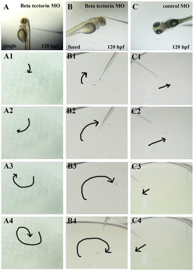
Abnormal swimming behaviors of β-tectorin morphants. β-Tectorin morphants were examined for their abilities to remain balance and react to a stimulus. Tactile stimulation was created by poking a zebrafish on the head with a glass tube: β-tectorin morphants with a single (panel A) and a fused otoliths (panel B), and a control with normal otoliths (panel C). Swimming behaviors of β-tectorin morphants at 5 days post-fertilization under stimulation were recorded with a digital video camera. β-Tectorin morphants with either single or fused otoliths failed to maintain their balance, tended to remain leaning on one side, remained on the bottom (panel A, B), and tended to swim in a corkscrew (panel A, A1 to A4) or circular manner (panel B, B1 to B4). Control zebrafish maintained their balance, had immediate responses to stimulation, and swam in a straight line (panel C, C1 to C4). The trails of the zebrafish movement were illustrated by dark arrows. PHENOTYPE:
|
|
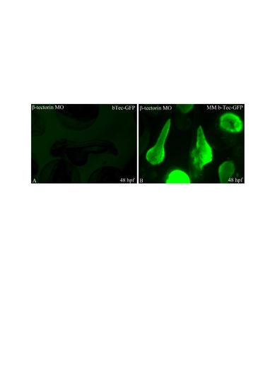
Control experiments for morpholino specificity. To determine the specificities of the morpholinos used, pCMV-GFP reporter plasmids containing a perfect (bTec-GFP) or mismatched (MM-b-Tec-GFP) MO target sequence were employed. Both bTec-GFP (A) and MM-b-Tec-GFP were co-injected with the β-tectorin MO. All images were taken from zebrafish embryos at 48 h post-fertilization. |
|
The morphology of β-tectorin morphants. The ATG MO injected zebrafish embryos with fused (a, b), and single otoliths (c, d) appeared to be normal without obvious defects. All photographs were taken at 72 hpf. |


