
- Title
-
Regulation of hub mRNA stability and translation by miR430 and the dead end protein promotes preferential expression in zebrafish primordial germ cells
- Authors
- Mickoleit, M., Banisch, T.U., and Raz, E.
- Source
- Full text @ Dev. Dyn.

Figure 2. hub mRNA is expressed in the head and in PGCs of zebrafish embryos and HuB protein is localized to perinuclear granules in the germ cells. A–C: Whole-mount in situ hybridization of wild type zebrafish embryos using an antisense probe directed against zebrafish hub RNA at the indicated developmental stages. hub mRNA is ubiquitously expressed in 10-hpf (A) and in 14-hpf embryos (B). In 24-hpf embryos, hub RNA is expressed in the head and in PGCs (inset) (C). D: The expression of nanos and hub mRNA determined by RT-PCR. Ovary cDNA, cDNA of embryos of the indicated developmental stages, and cDNA of germ cells isolated from 10hpf embryos were used as templates. E–H: Confocal microscopy images of a PGC in a 6hpf embryo showing the sub-cellular localization of the HuB-GFP fusion protein. The HuB fusion protein is localized to granular structures around the nucleus (E), which are co-labeled with the mCherry-Dnd fusion protein (F). The nucleus is marked by the CFP-histone protein (G) and the merged image is shown in H. |

Figure 3. hub translation is regulated by miR430. A: The level of the endogenous hub mRNA is increased in 10hpf MZdicer embryos compared to the level observed in wildtype embryos. As a comparison, qPCR analysis was performed to quantify nanos mRNA level in the same embryos. Error bars show SEM. ***P < 0.001 in t-test. B: Expression of GFP from mRNA containing the hub 32UTR is increased in the soma of MZdicer embryos (top right) as compared with the wild type control embryo (top left). mCherry fluorescent protein expressed from a co-injected mRNA that is not regulated by miRNA (mCherry-f-globin) was used as an internal control (bottom panels). Inset highlights the expression of GFP in the PGCs. C: The miR430 seed sequences within the hub 32UTR are labeled in blue and the mutations inactivating them in red. D–F: The predicted miR430 seed sequences in the hub 32UTR are functional in mediating translational repression. D: Expression of GFP from mRNA containing the wild type hub 32UTR (top left), compared to the expression from mRNA carrying mutations in the four predicted miR430 seeds in the 32 UTR (top right). Fluorescence intensity was measured in the soma (outline) at 16hpf and co-injected mCherry-F-globin served as internal control for the injection volume (bottom panels). E: A graph depicting the intensity of GFP expression measured for different mutated reporters as compared to that of wild type. n is the number of embryos analyzed and the error bars represent the SEM. ***P < 0.001 in t-test. F: Western blot of total protein extracted from 16-hpf embryos injected with RNA encoding GFP including the wild type or mutated hub 32UTR. EXPRESSION / LABELING:
|

Figure 4. Dnd counteracts the miR430-mediated inhibition of hub expression in PGCs. A: Dnd knockdown results in reduced expression of the Venus protein in PGCs of embryos injected with Venus.hub 32UTR RNA (top right) compared to the expression observed in control morpholino-treated embryos (top left). This effect is not observed for a CFP-based reporter containing the hub 32UTR with the mutated miR430 seed (compare bottom panels). B: Signal intensity measurements at 8hpf reveal a significantly reduced Venus expression. The signal is depicted as the ratio between the Venus and the CFP signals. n is the number of analyzed PGCs. Error bars show SEM. ***P < 0.001 in t-test. |
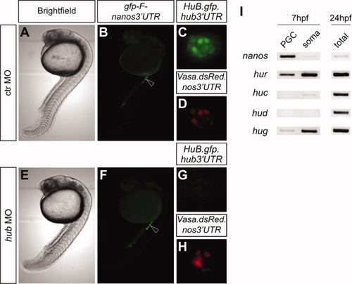
Figure 5. Knocking down HuB using morpholino antisense oligonucleotide does not affect the migration or survival of the PGCs. Embryos of transgenic fish expressing EGFP-F.nanos 32UTR were injected at one-cell stage either with a control morpholino (ctr MO, A–D), or with a morpholino inhibiting hub mRNA translation (hub MO, E–H) and analyzed at 24hpf. HuB knockdown embryos show no severe deformations in somatic development (compare A and E) and properly clustered PGCs in the region of the developing gonad (compare arrowheads B and F). In control morpholino-injected (ctr MO) embryos, the HuB-GFP reporter signal is detectable in perinuclear granules (C), whereas no signal is detected in embryos treated with hub MO (G) demonstrating sufficient morpholino-mediated inhibition. Both morpholinos had no effect on the level and localization of a Vasa.DsRed protein expressed in the same embryos. I: RT-PCR for Hu family members performed on sorted PGCs and somatic cells from 7hpf embryos. Similar to hub (Fig. 2D), hur and hug are expressed in PGCs. Amplification of nanos and the use of 24hpf total embryo cDNA serve as controls. |