
- Title
-
Core fucosylation is required for midline patterning during zebrafish development
- Authors
- Seth, A., Machingo, Q.J., Fritz, A., and Shur, B.D.
- Source
- Full text @ Dev. Dyn.
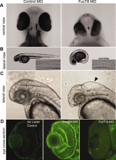
Knockdown of zfFucT8 leads to early midline defects. A: Ventral views of 48-hpf morphant embryos showing mild cyclopia (i.e., close-set eyes), and a smaller forebrain (arrow). B: Forty-eight-hpf FucT8 morphants have a curved body axis with U-shaped somites (inset), as well as (C) “swelling” in the hindbrain ventricle (arrowhead). D: AAL lectin stains α1,6 fucosyl residues in the eye, which is reduced to background levels in morpholino-injected embryos as compared to control-injected, or no lectin, controls. More than 500 embryos were injected with FucT8 MOs, of which 85-90% demonstrated the mutant phenotype. PHENOTYPE:
|
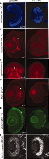
Defects in retinal cell differentiation in FucT8 morphant embryos. A:DAPI staining of eye cross-sections from 5-day-old control and FucT8 MO-injected embryos. Morphant embryos have a smaller retina, and although cell layers seem to be present, they are not as distinct as in control embryos. B,C: zn-5 immunostaining at 48 hpf identifies the RGCs (B, arrowhead) and optic chiasma (C, arrow) in control embryos, which are absent in morphant sections. One hundred percent of 20 injected embryos showed this phenotype. D: At 4 days, anti-CA-II antibody shows a lack of Müller glia (arrowhead) in the morphant retina. Eighty-five percent of 40 injected embryos showed this phenotype. E: zpr-1-positive photoreceptor cells are also severely reduced in FucT8 morphant eyes. Similar to 85% of 40 injected embryos showed this phenotype. F: BrdU incorporation illustrates increased cell proliferation in 28-hpf FucT8 morphant eyes. Eighty-two percent of 22 injected embryos showed this phenotype. |
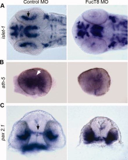
Patterning defects in the eye and forebrain of 27 hpf FucT8 morphant embryos. A: Ventral view of a FucT8 morphant embryo showing severe loss of islet-1 expressing retinal precursors (arrow). Ninety percent of 20 injected embryos showed this phenotype. B: Whole eyes showing a reduced number of ath-5-positive RGC precursors (arrow) in FucT8 morphant embryos. Similar to eighty-five percent of 40 injected embryos showed this phenotype. C: In situ hybridization using pax 2.1 shows that morphant embryos have a shorter and thicker optic stalk, relative to controls (arrow), and increased pax 2.1 expression in the eye field. Similar to eighty-five percent of 40 injected embryos showed this phenotype. |
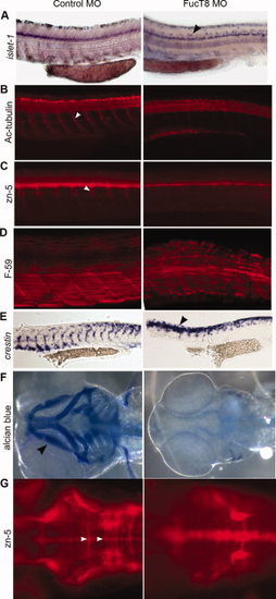
Defects in the trunk, spinal cord, and other regions of 48-hpf FucT8 morphants. A–E: Lateral views, anterior to the left. A:islet-1 expression at 48 hr in control and morphant embryos. In FucT8 morphants, motor neurons are disorganized and expanded in the dorso-ventral plane (arrowhead). Ninety percent of 20 injected embryos showed this phenotype. B: Immunostaining with anti-acetylated tubulin (Ac-tubulin) antibody shows a lack of motor neuron projections (arrowhead in Control MO) in the spinal cord of morphant embryos. Similar to eighty-five percent of 40 injected embryos showed this phenotype. C: zn-5 antibody labeling shows a loss of projections from the DRG (arrowhead) in FucT8 morphants. Ninety percent of 20 injected embryos showed this phenotype. D: FucT8 morphants have U-shaped somites with fewer slow muscle fibers, as shown by F-59 antibody. Ninety percent of 20 injected embryos showed this phenotype. E: In situ hybridization with crestin shows that neural crest cell migration is delayed in FucT8 morphants, remaining near their site of origin at the dorsal aspect of the neural tube (arrowhead). Similar to eighty-five percent of 40 injected embryos showed this phenotype. F: Ventral view of alcian blue–stained embryos show a lack of jaw cartilage (arrowheads) in FucT8 morpholino-injected embryos. One hundred percent of 4 injected embryos illustrate a similar phenotype. G: Dorsal views, anterior to the left. zn-5 staining shows that hindbrain commissures (arrowheads in Control MO) are significantly reduced in FucT8 morphants. Ninety percent of 20 injected embryos showed this phenotype. |
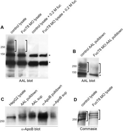
Identification of the major FucT8 substrates. A: AAL lectin blot of control and FucT8 morphant embryo lysates resolved by SDS-PAGE. The morphant lysate has reduced AAL-positive proteins in the high molecular weight region (brackets). AAL lectin binding to these high molecular weight polypeptides is inhibited by excess fucose in both control and morphants samples, whereas the lower two bands are not and represent non-specific bands (indicated by the asterisk). B: Control and FucT8 morphant lysates were incubated with AAL-conjugated beads to precipitate the core fucosylated proteins, which were subsequently resolved by SDS-PAGE and subjected to AAL lectin blotting. AAL beads precipitate high molecular weight AAL-reactive glycoproteins that are not present in similarly treated morphant lysates (brackets). *, The same non-specific protein band seen in A. C: AAL-conjugated beads were used to pull down core fucosylated proteins from a HepG2 cell lysate, which were resolved by SDS-PAGE and probed with ApoB antibody. The anti-ApoB detects a large molecular mass polypeptide in HepG2 lysates that migrates above the 250-kDa marker, and anti-ApoB antibodies were able to immunoprecipitate a polypeptide of similar size. AAL beads were also able to precipitate an ApoB-reactive polypeptide of similar molecular weight, although not all ApoB was precipitated by AAL, since ApoB-reactive material remained in the AAL bead supernatant. D: Coomassie blue–stained gel of high molecular weight polypeptides, which correspond to the ApoB-containing, AAL-reactive bands in control embryos. The amount of high molecular weight ApoB polypeptides (brackets) appears to be somewhat reduced in FucT8 morphants. PHENOTYPE:
|
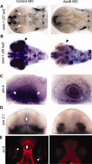
Knockdown of zfApoB phenocopies FucT8 morphants. A,B:islet-1 expression in the optic field is severely reduced (arrowheads) in ApoB morphants, similar to that seen in FucT8 morphants (see Fig. 4A) (27 hpf: 86% of 50 injected embryos showed this phenotype; 48 hpf: 85% of 40 injected embryos showed this phenotype). C: Whole eyes, dissected out from ath-5-labeled embryos, shows a reduced number of ath-5-expressing cells (arrowheads) in 27-hpf ApoB morphants, as seen in FucT8 morphants (see Fig. 4B). Eighty-six percent of 43 injected embryos showed this phenotype. D:pax 2.1 expression in 27-hpf ApoB morphants illustrates a shorter and thicker optic stalk, relative to controls (arrow), and parallels what is seen in FucT8 morphants (see Fig. 4C). Eighty-two percent of 49 injected embryos showed this phenotype. E: Ventral views, anterior to the top. Immunostaining with zn-5 antibody shows loss of RGCs (arrowheads) and optic chiasma (arrow) in 48-hpf ApoB morphant embryos phenocopying that seen in FucT8 morphants (see Fig. 3C). Ninety-eight percent of 47 injected embryos showed this phenotype. |
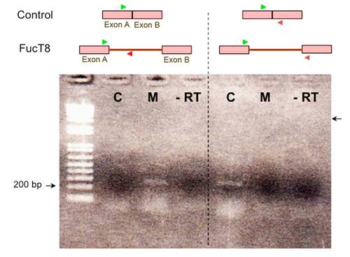
Supporting Figure S1: RT-PCR analysis indicates that FucT8 morpholino oligonucleotides were effective in blocking processing of FucT8 mRNA. RNA extracted from 24-hpf control and FucT8 morphant embryos was subjected to RT-PCR using two primers pairs; the first pair (green, red arrows) produced a 270-bp fragment (arrow) in FucT8 morphants, that is not present in control-injected embryos. The second primer pair used the same forward primer (green) along with a reverse primer (blue) that generated a 266-bp product in control embryos, as well as a faint, larger PCR product of 2,858 bp (arrow) in FucT8 morphants that is not easily reproduced in the figure, as the PCR reaction was optimized for smaller reaction products. |
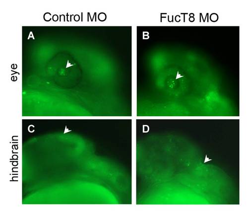
Supporting Figure S2: Cell death is slightly increased in FucT8 morphants. Apoptotic cells (arrowheads) were assayed at 28 hpf by Acridine Orange staining as described in the Experimental Procedures section. Representative views of forebrain/eye (A,B) and hindbrain (C,D) are shown. Eighty percent of 34 injected embryos showed a slight generalized increase in cell death. |
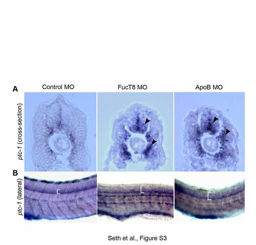
Supporting Figure S3: Shh signaling is affected in FucT8 and ApoB morphants. Sections of 24-hpf embryos subjected to in situ hybridization for ptc-1. A: In both FucT8 and ApoB morphants, there is an increase in ptc-1 expressing cells around the notochord and in the spinal cord (arrowheads). B: Lateral views of ptc-1 expression as seen by whole mount in situ hybridization of 24 hpf embryos. ptc-1-expressing cells appear to be increased and more dispersed (brackets) in FucT8 and ApoB morphants. Ninety percent of 10 injected embryos illustrated this phenotype. |

Unillustrated author statements PHENOTYPE:
|