
- Title
-
Separating early sensory neuron and blood vessel patterning
- Authors
- Miller, L.C., Freter, S., Liu, F., Taylor, J.S., Patient, R., and Begbie, J.
- Source
- Full text @ Dev. Dyn.
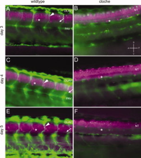
Loss of blood vessels in the cloche mutant zebrafish does not affect the positioning of the DRG. A,C,E: Lateral view of fli1:gfp transgenic embryos stained for Elavl3/4 at 3 (A), 4 (C), and 5 (E) dpf. B,D,F: Lateral view of tg(fli1:gfp) cloche mutant embryos stained for Elavl3/4 at 3 (B), 4 (D), and 5 (F) dpf. The fli1:gfp labelling indicates the vasculature and in the normal embryos the intersomitic vessels (arrowhead), dorsal longitudinal anastomotic vessel (DLAV), parachordal vessel (PAV), and vertebral artery (arrow) can be seen, whereas these are all absent in the cloche embryos. Elavl3/4-positive neurons can be seen in the neural tube (NT) and in the developing DRG (*). The dorsoventral position of the DRG is the same in both normal and mutant embryos. |