- Title
-
Atoh8, a bHLH transcription factor, is required for the development of retina and skeletal muscle in zebrafish
- Authors
- Yao, J., Zhou, J., Liu, Q., Lu, D., Wang, L., Qiao, X., and Jia, W.
- Source
- Full text @ PLoS One
|
Expression patterns of the zebrafish atoh8 gene during morphogenesis. Lateral view, anterior to the left (A,C,F–K). Top view (B,D). Dorsal view, anterior to the left (E). (A) 70% epiboly. (B) 6-somite. (C–E) 15-somite. (F) 17-somite. (G) 21-somite.(H,M,N) 24 hpf. (I) 36 hpf. (J) 48 hpf. (K) 72 hpf. (L–M)horizontal section of retina from 15-somite to 17-Somite stage embryos. Abbreviations: b, brain; hb, hind brain; lp, lens primordium;ncc,neuron crest cell; op, optic vesicle; re, retina; sm, somite. EXPRESSION / LABELING:
|
|
Phenotypes of translation blocking morpholino (MO1) knockdown embryos. (A–B)13-somite stage. (C–E) 24 hpf. (F–H) 48 hpf. (I–J) 72 hpf. (K–N) somite of mylz2-GFP transgenic zebrafish embryos at 48 hpf. (O–P) mylz2-GFP transgenic zebrafish embryos at 7dpf. A, C–D, F–G, I, K, M and O are Atoh8-MO treated embryos: A, C and F show severe abnormality, D, G, I, K, M and O show mild abnormality, and B, E, H, J, L, N and P are control-MO treated embryos. Atoh8-MO-treated embryos show shrunken eyes and head, a curved body axis with indistinct somite boundaries and yolk extension, accompanied by a U-shaped somite formation. In addition, the branchial arches are greatly reduced(O–P). In all panels but P, the dorsal view is up and the anterior is to the left. P shows the ventral view and the anterior to the left. Abbreviations: re, retina; sm, somite; ba, branchial arches. EXPRESSION / LABELING:
PHENOTYPE:
|
|
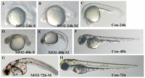
Phenotypes of splicing blocking morpholino (MO2) knockdown embryos. (A–C) 24 hpf. (D–F) 48 hpf. (G–H) 72 hpf. A–B, D–E and G are MO2 treated embryos: A and D, severe abnormality, B, E and G, mild abnormality and C, F and H, control-MO treated embryos. The figure shows that splicing blocking morpholino treated embryos show similar phenotypes as that of translation blocking MO1-treated ones. PHENOTYPE:
|
|
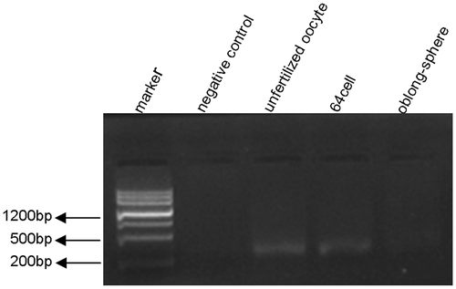
RT- PCR detection of maternally supplied atoh8 mRNA in zebrafish. Maternally supplied athoh8 mRNA was detected by RT-PCR in both unfertilized oocytes and 64-cell stage but significantly reduced in oblong-spheres. The negative control contains no sample template. |
|
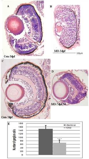
Transverse sections of Atoh8-MO treated embryo retina. (A,C) Control eyes at 3dpf and 7dpf, respectively. (B,D) Eyes of MO-treated fish at 3dpf and 7dpf, respectively. The section shows that the retina did not develop normal neuronal stratification compared with the control at 3dpf. By 7pdf, lamination had formed in the treated embryos, however, the retinal size was remarkably reduced (D), and the number of retinal ganglion neurons was significantly reduced in MO fish (E). (p<0.001). Besides, the photoreceptor layer of the morphant was less elongated. In all panels, the ventral is down. PHENOTYPE:
|
|
TUNEL assay of retina of Atoh8 knockdown embryos at 48hpf. (A) severely defective embryo retina shows serious cell apoptosis and (B) a mildly malformed embryo retina appears to contain only a minor population of apoptotic cells compared to the control (C). The brown cells were TUNEL-positive (arrow). PHENOTYPE:
|
|
Retinal development is disrupted in Atoh8 knockdown embryos. (A–B) 15-Somite stage; (C–D,G–H) 48 hpf; (E–F) 36 hpf, (A,C,E,G) morphant and (B,D,F,H) control. Staining of pax6a showed that retinal region was remarkably reduced (arrow) in this MO-treated embryo. Atoh5 expression was present throughout most of the neural retinal at 36 hpf in the control embryo(E) while present only in dorsal and temporal retina in the MO-treated embryo(F). By 48 hpf, atoh5 localized throughout retina ganglion cells in the control(G) but present only in ventral retina in the MO-treated embryo(H). In addition, the signals of these two genes were much weaker than those of the control, indicating that knock-down of Atoh8 may have influenced retinal organization. In all panels, the dorsal side is up and the anterior is to the left. EXPRESSION / LABELING:
|
|
Skeletal muscle development is altered in Atoh8 knock-down embryos. (A–H) myoD in situ hybridization; (I–L) myogenin in situ hybridization. (A–C) 9-Somite stage; (D–E) 15-Somite stage; (G–H) 36 hpf; (C,F,H,J and L) are controls and the remainder are morphants with (B) and (E) the severely affected embryos. Expression of the skeletal marker myoD in paraxial mesoderm showed to tendency towards a ventral localization (A,D) or the boundaries of the posterior somites were indistinct(B,C) and a U-shaped somite formation appeared in the morphant (G). Expression of the skeletal marker myogenin provides further evidence of abnormality (I–L). EXPRESSION / LABELING:
PHENOTYPE:
|
|
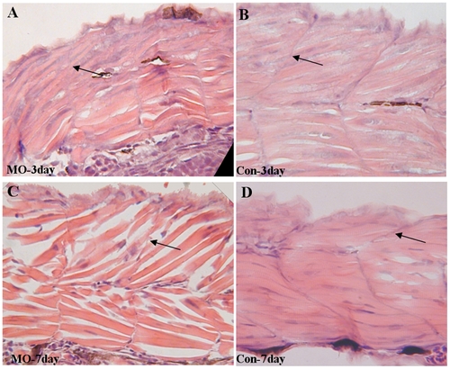
Absence of Atoh8 affects the myogenic differentiation of skeletal muscle. (A–B) transverse section of somites at 3dpf and (C–D) transferse section of somites at 7dpf. (A,C) somites of Atoh8-treated embryos; (B,D) the related controls. Note that the transverse myoseptum of MO-treated embryos is ill-defined and incomplete and that the somites form a U-shape or even appear cuboidal. Somites in controls, on the other hand, are chevron shaped with a distinct somite boundary. In addition, differentiated muscle fibers in myotomes show a less compact and orderly looking arrangement in the morphant. PHENOTYPE:
|


