- Title
-
Expression of the zebrafish CD133/prominin1 genes in cellular proliferation zones in the embryonic central nervous system and sensory organs
- Authors
- McGrail, M., Batz, L., Noack, K., Pandey, S., Huang, Y., Gu, X., and Essner, J.J.
- Source
- Full text @ Dev. Dyn.
|
Identification and expression analysis of zebrafish CD133/prominin1 paralogues prom1a and prom1b. A: Phylogenetic analysis of zebrafish prominin genes. aga, Anopheles gambiae; can, Canis familiaris; cel, Caenorhabditis elegans; dme, Drosophila melanogaster; dre, Danio rerio; gga, Gallus gallus; hsa, Homo sapiens; mus, Mus musculus; pan, Pan troglodytes; rno, rtn, Rattus norvegicus; xtr, Xenopus tropicalis. B: RT-PCR showing relative levels of prom1a and prom1b expression throughout development and in adult tissues. Control, expression of ribosomal protein S6 kinase b, polypeptide 1, (rps6kb1). |
|
Expression of zebrafish prom1a and prom1b in developing sensory organs and CNS of 24- and 36hpf embryos. A: Lateral view, 24hpf. prom1a expression in the lens anlage, epiphysis, and otic vesicle. B: Lateral view, 24hpf. prom1b expression in the epiphysis (arrowhead). C: Lateral view, 36hpf. prom1a expression in the ventricular zone along the dorsal surface of the midbrain tegmentum and extending into the hindbrain. D: Camera lucida drawing of 35hpf zebrafish embryo (Kimmel et al.,[1995]) showing position of cross-sections shown in E and F. E: At 36hpf, prom1a expression in the ventricular zone of the midbrain tegmentum and lens fiber cells (inset). F: At 36hpf, prom1b expression in the epiphysis. G: Dorsal view, 36hpf. prom1a expression extends from the epiphysis posteriorly along the tectum (arrowhead). H: Dorsal view, 36hpf. prom1b expression is restricted to the epiphysis. ant, anterior; cb, cerebellum; ea, eye anlage; ep, epiphysis; ey, eye; h, hypothalamus; hb, hindbrain; le, lens epithelium; lf, lens fiber cells; mt, midbrain tegmentum; op, olfactory placode; ot, optic tectum; ov, otic vesicle; pos, posterior; tc, telencephalon; th, thalamus; vz, ventricular zone. Scale bars = 100 μm. |
|
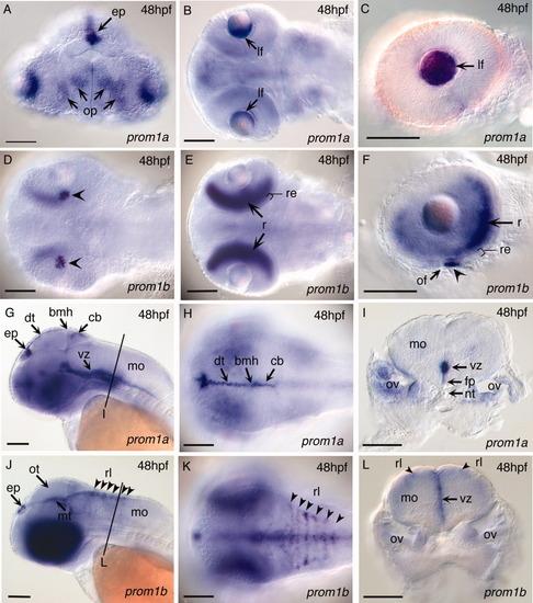
Expression of zebrafish prom1a and prom1b in the sensory organs and CNS of the zebrafish embryo at 48hpf. prom1a (A-C, G-I) and prom1b (D-F, J-L) expression. Sensory organs: prom1a expression in the epiphysis (A, G), olfactory epithelium of the olfactory placodes (A), and lens epithelium of the eye (B, C). prom1b expression in the epipyhysis (J), retinal epithelium (D, F, arrowheads), and marginal layer of the neural retina in the eye (E, F). CNS: G-I: prom1a expression in the tectum and ventricular zones of the hindbrain. J-L: prom1b expression in the midbrain tegmentum and ventricular zones of the hindbrain. A: Anterior frontal view; B, E: dorsal medial optical section; D: ventral view; G, J: lateral view; H, K: dorsal view. I, L: cross-section through the hindbrain neural tube. bmh, boundary between mid- and hindbrain; cb, cerebellum; dt, dorsal optic tectum; ep, epiphysis; fp, floor plate; lf, lens fiber cells; mo, medulla oblongata; mt, midbrain tegmentum; nt, notochord; of, optic fissure; op, olfactory placode; ot, optic tectum; ov, otic vesicle; r, neural retina; re, retinanl epithelium; rl, rhombic lip; vz, ventricular zone. Scale bars = 100 μm. EXPRESSION / LABELING:
|
|
Increased expression of prom1a and prom1b in epitheloid-like cells in tp53M214K/tp53M214K induced neoplasm. A, B, E, F: Gross morphology and histopathology of abdominal (A, B) and ocular (E, F) neoplasms in 12-month-old adult homozygous tp53M214K/tp53M214K zebrafish. B: Histopathological features of the abdominal neoplasm show regions with predominantly spindle cells (solid line box) or predominantly epitheloid cells (dashed line box), consistent with a diagnosis of zMPNST (Berghmans et al., [2005]). F: Histopathological features of ocular tumor show predominantly spindle cells, consistent with a diagnosis of zMPNST (Berghmans, et al., 2005). C, D: Expression of prom1a (C) and prom1b (D) is detected within epitheloid-like cells in the abdominal neoplasm (solid line box), but is absent from the region containing predominantly spindle cells (dashed line box). G, H: Absence of expression of prom1a (G) and prom1b (H) in spindle cells of the ocular neoplasm. Scale bars in A, E = 0.5 cm; in H = 50 μm. EXPRESSION / LABELING:
|
|
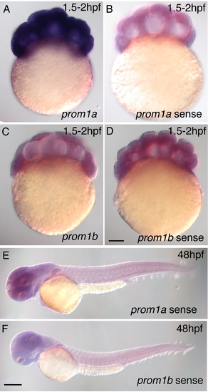
Control experiments to demonstrate specificity of the prom1a and prom1b antisense probes used for in situ hybridization. Specific labelling using antisense probes (A, C) versus background signal in embryos hybridized with sense probes (B, D-F). Intense labelling with the prom1a probe in the 2hpf embryo (A) is consistent with RT-PCR demonstrating high levels of maternal message within the 1hpf embryo. In contrast, the absence of specific labeling by prom1b in the 2hpf embryo (C) is consistent with RT-PCR showing barely detectable levels of maternal message in the 1hpf embryo. Figure 1B, lane 1, compare prom1a and prom1b. Scale bars = 100 μm. |


