
- Title
-
Use of Zebrafish to Probe the Divergent Virulence Potentials and Toxin Requirements of Extraintestinal Pathogenic Escherichia coli
- Authors
- Wiles, T.J., Bower, J.M., Redd, M.J., and Mulvey, M.A.
- Source
- Full text @ PLoS Pathog.

Phagocyte recruitment and abatement within the zebrafish pericardial cavity. (A and B) Zebrafish embryos were inoculated via the P.C. with 4,000–6,500 CFU of (A) the K12 strain MG1655 or (B) the ExPEC isolate F11, both carrying pGEN-GFP(LVA) for constitutive expression of destabilized GFP protein (green). Samples were fixed at 0, 6, and 24 hpi and processed for fluorescent confocal microscopy. Phagocytes (red) were detected using L-plastin-specific antibody. For clarity, the merged image at each time point is accompanied by images showing only corresponding single channel signals from bacteria or L-plastin. Each embryo was visualized by stitching together two z-projections generated from 40 to 50 5-μm-thick optical sections. Scale bar = 200 μm. (C) Tg(mpo::GFP) zebrafish embryos, in which GFP expression is under control of the neutrophil-specific MPO promoter, were injected via the P.C. with ∼5,000 CFU of the ExPEC isolate F11. At 4 hpi, samples were fixed and stained using anti-L-plastin antibody to label total phagocytes (red) relative to the GFP-positive neutrophils (green plus red). Bacteria present within the P.C. are not shown. Scale bar = 100 μm. (D) Diagram of a 48 hpf embryo with the area imaged in (C) highlighted by a red box. Embryos shown here are representative. All fish were viable at the time of sacrifice, except the 24 hpi F11-infected fish, which died prior to collection. |

Phagocyte localization and bacterial internalization within the pericardial cavity. Zebrafish embryos were injected via the P.C. with 4,000–6,500 CFU of (A–C) wt UTI89, (D–F) UTI89ΔhlyA, or (G–I) UTI89Δcnf1, each carrying pGEN-GFP(LVA) for constitutive expression of destabilized GFP protein (green). At 6 hpi, samples were fixed and processed for fluorescent confocal microscopy, using anti-L-plastin antibody to label phagocytes (red). Examples of internalized (solid arrowheads) and free extracellular (hollow arrowheads) bacteria in (A, D, and G) are shown at higher magnification in (B, E, and H) and (C, F, and I), respectively. Representative images are shown. All fish were viable at the time of sacrifice. Scale bars = 100 μm. (J) Diagram of a 48 hpf embryo with the area imaged in (A), (D), and (G) highlighted by a red box. |
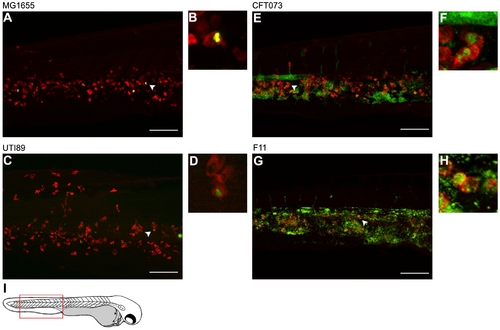
Differential growth and phagocytosis of E. coli isolates within the blood. Zebrafish embryos were infected via the blood with 4,000–6,500 CFU of (A and B) MG1655, (C and D) UTI89, (E and F) CFT073, or (G and H) F11. All bacterial strains carry pGEN-GFP(LVA) for constitutive expression of destabilized GFP (green). At 12 hpi, samples were fixed and phagocytes (red) were labeled using L-plastin-specific antibody for visualization by fluorescent confocal microscopy. Regions highlighted by arrowheads in (A), (C), (E), and (G) are shown further magnified in panels (B), (D), (F), and (H), respectively. All images shown are representative of the pool of embryos imaged. MG1655- and UTI89-infected embryos were viable and healthy in appearance prior to sacrifice for microscopy, whereas fish inoculated with CFT073 or F11 were notably sick and near death at time of collection. Scale bars = 100 μm. (I) Diagram of a 48 hpf embryo with the region imaged in (A), (C), (E), and (G) denoted by a red box. |
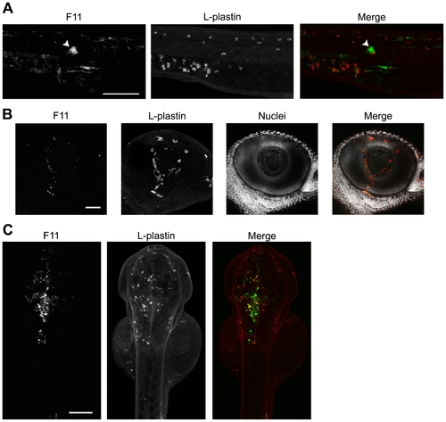
ExPEC isolates capable of blood colonization can disseminate and persist in a variety of host microenvironments. F11, carrying pGEN-GFP(LVA) for constitutive expression of destabilized GFP (green), was inoculated into the blood using a dose of 4,000–6,500 CFU. Samples were fixed at 6 or 12 hpi and phagocytes (red) were labeled using L-plastin-specific antibody for visualization by fluorescent confocal microscopy. (A) 20X z-projection of the tail region from an F11-infected embryo at 12 hpi. The arrowhead indicates a commonly observed bacterial microcluster. (B) 40X z-projection of the eye from an embryo at 6 hpi with F11. Hoechst nuclear dye (grey) was used to highlight the anatomical structure of the eye. (C) Dorsal to ventral 10X z-projection of the head region of an embryo at 12 hpi with F11. Scale bars = 100 μm for (A) and (C) and 50 μm for (B). |