- Title
-
Domain-specific regulation of foxP2 CNS expression by lef1
- Authors
- Bonkowsky, J.L., Wang, X., Fujimoto, E., Lee, J.E., Chien, C.B., and Dorsky, R.I.
- Source
- Full text @ BMC Dev. Biol.
|
Sequential expression of foxP2 and lef1 in the CNS during embryogenesis. Whole-mount in situs for lef1 (A-C) and foxP2 (D-F) at 24 hpf, 30 hpf, and 36 hpf. Lateral views, anterior to left, dorsal up; eyes have been removed to facilitate visualization. Scale bar = 50 μm. (Abbreviations: dd, dorsal diencephalon; hb, hindbrain; hy, hypothalamus; mhb, mid-hindbrain boundary; tec, tectum; tel, telencephalon.) (A-C): lef1 is expressed in the hypothalamus, dorsal midbrain, and MHB at 24 hpf, with expression extending to the tectum at 30 hpf. By 36 hpf expression is confined primarily to the hypothalamus and tectum. (D-F): foxP2 is expressed in the tectum and MHB starting at 36 hpf. Earlier expression (24 hpf and 30 hpf) is confined to the telencephalon. |
|
Co-expression of foxP2 and lef1 in the tectum. Whole-mount double in situ confocal imaging for foxP2 and lef1 at 36 hpf, dorsal views, anterior up. Abbreviations: hb, hindbrain; tec, tectum; tel, telencephalon. (A) Z-stack projection of foxP2 (green) overlaid on brightfield image of lef1 expression in the tectum. The region shown in higher magnification in (B-D) is boxed in yellow. Scale bar = 50 μm. (B-D) Single optical plane showing lef1 (red- BM Purple), foxP2 (green- Alexa 488), and co-expression in the tectum. Arrow points to a representative co-expressing cell. Scale bar = 25 μm. EXPRESSION / LABELING:
|
|
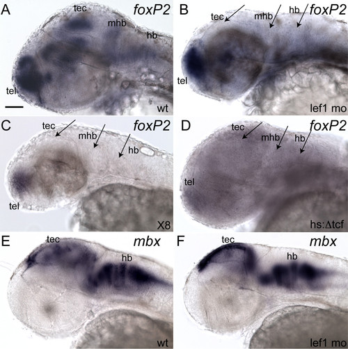
Loss of lef1 leads to absent foxP2 expression in the tectum, mid-hindbrain boundary, and hindbrain. Whole-mount in situs at 36 hpf; anterior to left, dorsal up, eyes removed. Scale bar = 50 μm. (Conditions: hs:Δtcf, Tg(hsp70l:Δtcf-GFP)w26; lef1 mo, lef1 morphants; wt, wild type; x8, homozygous Df(LG01)x8. Abbreviations: hb, hindbrain; mhb, mid-hindbrain boundary; tec, tectum; tel, telencephalon.) (A, B) lef1 morphant (B) lacks expression in tectum, MHB, and hindbrain (arrows), compared to wild-type (A). (C) Df(LG01)x8 homozygote lacks expression in tectum, MHB, and hindbrain (arrows). (D) Tg(hsp70l:Δtcf:GFP)w26 embryo, three hours post-heat shock, lacks expression in tectum, MHB, and hindbrain (arrows). (E, F) mbx in situs; staining in tectum and hindbrain is indistinguishable between wild type and lef1 morphants (F). EXPRESSION / LABELING:
|
|
Confocal live whole-mount images of foxP2 enhancers. Pictures show GFP expression at 72 hpf (except foxP2-enhancerD, taken at 48 hpf). The eye has been removed in panels E and G to facilitate visualization. Scale bar = 100 μm. (A, C, E, G, I): lateral views, anterior left, dorsal up. (B, D, F, H, J): dorsal views, anterior up. (Abbreviations: di, diencephalon; hb, hindbrain; tec, tectum; tel, telencephalon; arrows, GFP-expressing cells in the eye. (A, B) Tg(foxP2-enhancerA:EGFP)zc42; (C, D) Tg(foxP2-enhancerA.1:EGFP)zc44; (E, F) Tg(foxP2-enhancerA.2:EGFP)zc46; (G, H) Tg(foxP-enhancerB:EGFP)zc41; (I, J) Tg(foxP2-enhancerD:EGFP)zc47. EXPRESSION / LABELING:
|
|
lef1 is necessary for expression from foxP2-enhancerA.1, foxP2-enhancerB, and foxP2-enhancerD. Whole-mount gfp in situs at 36 hpf; anterior to left, dorsal up, eyes removed. Scale bar = 50 μm. Conditions (wild type, wt; or lef1 morphant, lef1 MO) are shown above the panels, enhancer names to left. (Abbreviations: dd, dorsal diencephalon; hb, hindbrain; tec, tectum; tel, telencephalon.) (A) Tg(foxP2-enhancerA.1:EGFP)zc44 expresses in telencephalon, tectum, and hindbrain. (B) Tg(foxP2-enhancerA.1:EGFP)zc44 embryo injected with lef1 morpholino lacks GFP expression in tectum and hindbrain (arrows), although telencephalic expression persists. (C) Tg(foxP2-enhancerB:EGFP)zc41 expresses in telencephalon and hindbrain. (D) Tg(foxP2-enhancerB:EGFP)zc41 embryo injected with lef1 morpholino lacks GFP expression in the hindbrain (arrow), but telencephalic expression is present. (E) Tg(foxP2-enhancerD:EGFP)zc47 expresses in dorsal diencephalon and tectum. (F) Tg(foxP2-enhancerD:EGFP)zc47 embryo injected with lef1 morpholino lacks GFP expression in the tectum (arrow), but dorsal diencephalic expression persists. (G) Tg(elavl3:EGFP)zf8 embryo shows GFP expression in all post-mitotic neurons. (H) Tg(elavl3:EGFP)zf8 embryo injected with lef1 morpholino still has GFP expression in the tectum and hindbrain. EXPRESSION / LABELING:
|
|
ChIP analysis of foxP2-enhancerA.1 and foxP2-enhancerB genomic regions at 30 hpf. (A) Diagram of foxP2-enhancerA.1 and foxP2-enhancerB regions. PCR amplicon locations and names are indicated above the genomic region; predicted Lef1 binding sites are shown as ovals. (B) Agarose gel analysis of ChIP PCR for foxP2-enhancerA.1, showing the PCR products for FP2700, FP300, and B3end. In wild type (wt) embryos, ChIP shows significant enrichment of the FP2700 product and slight enrichment of the FP300 product, compared to both the no antibody (Ab) control, and the ChIP of homozygous Df(LG01)x8 embryos (Mock: water control for the PCR reaction.) In contrast, the B3end product showed no enrichment relative to the Df(LG01)x8 embryos. (C) Agarose gel analysis of ChIP PCR for foxP2-enhancerB, showing the FPb2600 PCR product. In wt embryos, ChIP shows significant enrichment of the FPb2600 product, compared to both the no antibody and homozygous Df(LG01)x8 controls. |
|
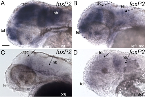
Loss of lef1 leads to absent foxP2 expression in the tectum, mid-hindbrain boundary, and hindbrain. Whole-mount in situs at 48 hpf; anterior to left, dorsal up, eyes removed. Scale bar = 50 μm. (Conditions: hs:Δtcf- Tg(hsp70l:Δtcf:GFP)w26, lef1 mo- lef1 morphants, wt- wild type, x8- homozygous Df(LG01)x8. Abbreviations: hb: hindbrain; mhb: mid-hindbrain boundary; tec: tectum; tel: telencephalon.) (A, B) lef1 morphant (B) lacks expression in tectum and has diminished expression in the hindbrain (arrows), compared to wild-type (A). (C) In Df(LG01)x8 homozygote, expression is absent in the tectum and hindbrain (arrows). (D) In Tg(hsp70l:Δtcf:GFP)w26 embryo, collected four hours after heat shocking, expression is absent from the tectum and hindbrain (arrows). MHB expression is reduced but not absent, perhaps because lef1 induction of foxP2 expression is already sufficiently established by the time of heat-shock. EXPRESSION / LABELING:
|
|
lef1 is necessary for expression from foxP2-enhancerA.1, foxP2-enhancerB, and foxP2-enhancerD. Whole-mount confocal images at 52 hpf (except foxP2-enhancerD), lateral view, anterior left, dorsal up. Conditions (wild type- wt, or lef1 morphant- lef1 MO) are shown above the panels, while the enhancer name is to the left of the panels. (Abbreviations: hb: hindbrain; tec: tectum; tel: telencephalon.) (A) Tg(foxP2-enhancerA.1:EGFP)zc44 expresses in telencephalon, tectum, and hindbrain. (B) Tg(foxP2-enhancerA.1:EGFP)zc44 embryo injected with lef1 morpholino: GFP expression is absent in the tectum and hindbrain (arrows), although telencephalic expression persists. (C) Tg(foxP2-enhancerB:EGFP)zc41 expresses in telencephalon, tectum, and hindbrain. (D) Tg(foxP2-enhancerB:EGFP)zc41 embryo injected with lef1 morpholino: GFP expression is absent in the hindbrain (arrow), but still present in the telencephalon. (E) Tg(foxP2-enhancerD:EGFP)zc47 expresses in tectum. (F) Tg(foxP2-enhancerD:EGFP)zc47 embryo injected with lef1 morpholino: GFP expression is absent in the tectum (arrow). (G) Tg(elavl3:EGFP)zf8 embryo shows GFP expression in all post-mitotic neurons. (H) Tg(elavl3:EGFP)zf8 embryo injected with lef1 morpholino: GFP expression is still present in the tectum and hindbrain. (I) Tg(pax2a:GFP)e1 embryo shows expression in the MHB and hindbrain. (J) Tg(pax2a:GFP)e1 embryo injected with lef1 morpholino has persistent expression in the MHB and hindbrain. EXPRESSION / LABELING:
|


