
- Title
-
miR-145 directs intestinal maturation in zebrafish
- Authors
- Zeng, L., Carter, A.D., and Childs, S.J.
- Source
- Full text @ Proc. Natl. Acad. Sci. USA

miR-145 expression pattern and loss of function phenotype. (A) Lateral view of whole-mount in situ expression of miR-145 at 16–19S shows ubiquitous expression. (B) Later at 96 hpf, miR-145 is expressed in the gut (g) and swimbladder (sb) in wild-type embryos. (C) Longitudinal section of 96 hpf embryo shows miR-145 expressed strongly in the gut smooth muscle cells (white arrowhead) and weakly in gut epithelium. (D–F) Animals injected with miR-145 MO have hydrocephalus in the hind brain ventricle (*) at 48 hpf. (G–I) At 96 hpf, miR-145 morphants have severe pericardial edema (black arrowhead), and an underdeveloped gut and swimbladder (black arrow). In situ hybridization staining of αsma (J–L) and sm22α-b (M–O) in 96 hpf embryos shows an increase of sm22α-b expression but no change of αsma in the miR-145 morphant gut. Gut morphology in the miR-145 morphant is unlooped as compared to the broader tube-like morphology found in UIC and miR-145 control MO embryos. Arrows mark the gut. (P) qPCR of αsma and sm22α-b in miR-145 morphants at 48 hpf. (Q and R) Histology of 96 hpf zebrafish gut. miR-145 morphant shows a thinner layer of SMCs (SM) and lack of villi in the epithelial layer as compared to UIC embryos. (Scale bars, 200 μm except for C, Q, and R, which are 50 μm.) EXPRESSION / LABELING:
|
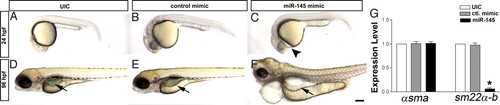
miR-145 overexpression affects gut and heart development. (A–C) Overexpression of miR-145 causes mild pericardial edema (arrowhead) in 24 hpf embryos, (D–F) which then develops into severe pericardial edema accompanied by a unlooped gut at 96 hpf. The control mimic RNA injection has no effect. (G) Overexpression of miR-145 leads to strong down-regulation of sm22α-b but does not affect αsma levels in 48 hpf embryos as measured by qPCR. Arrows, gut. (Scale bar, 200 μm.) |
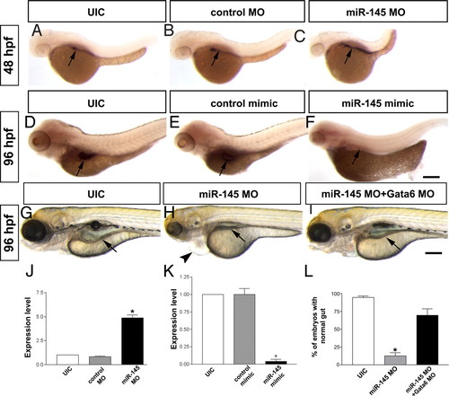
Normalization of gata6 levels rescues loss of miR-145. (A–C) Knockdown of miR-145 results in increased expression of gata6 in zebrafish gut at 48 hpf. (D–F) Overexpression of miR-145 leads to decreased gata6 expression in the gut of 96 hpf embryos. Pericardial edema (arrowhead) is also found in miR-145 morphant. (G–I) gata6 knockdown normalizes the gut morphology (arrow) of miR-145 morphants. (J) qPCR shows strong up-regulation of gata6 in miR-145 morphants of 48 hpf. (K) qPCR indicates significantly decreased expression of gata6 in 48 hpf miR-145 mimic treated embryos. Arrows, gut; bars, mean ± SEM. *, P < 0.01. (L) The number of embryos exhibiting normal gut morphology in the rescue was analyzed by using Student′s t test (P < 0.05, n = 286). (Scale bar, 200 μm.) |

Phenotype after injection of miR-145 morpholino and mimic. (A) LNA in situ hybridization shows ubiquitous expression of miR-145 in uninjected wild-type embryos (UIC) and B) control MO-injected embryos. (C) miR-145 expression is significantly reduced by miR-145 MO and (D) pre-miR-145 morpholino. (E) Expression of miR-145 in uninjected or (F) control mimic expressing embryos is low in comparison to (G) miR-145 mimic-injected embryos. (H) qPCR indicates significant knocking down of miR-145 in 24 hpf embryos treated with mature miR-145 and pre-miR-145 morpholino. (I–K) Co-injection of miR-145 mimic with miR-145 morpholino can partially rescue the phenotype of miR-145 morphants. (L) pre-miR-145 MO induces a similar phenotype to miR-145 MO with gut morphological defects (arrow) and pericardial edema (arrowhead) in 96 hpf embryos. Bars, mean ± SEM. *, P < 0.05. (Scale bars, 200 μm.) PHENOTYPE:
|
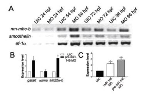
Loss of miR-145 leads to smooth muscle marker expression changes. (A) RT-PCR shows increased expression of nm-mhc-b and smoothelin in miR-145 morphants at 54, 72, and 96 hpf in comparison with the housekeeping gene EF1α. (B) qPCR indicates significantly increased expression of gata6 and sm22α-b but similar level of αsma in 48 hpf embryos treated with pre-miR-145 morpholino. (C) qPCR shows the increased expression of nm-mhc-b in miR-145 MO or pre-miR-145 morpholino treated embryos at 48 hpf. Bars, mean ± SEM. *, P < 0.05. |

Injection of the Gata6-TPmiR145 target protector morpholino mimics the miR-145 morphant phenotype. (A and B) Gata6-TPmiR145 target protectorinjected embryos display gut morphological defects and pericardial edema (arrowhead) at 96 hpf as compared to uninjected wild-type embryos. Expression of gata6 (C and D) and sm22α-b (E and F) is increased in Gata-6-TPmiR145 morphants. Arrows, gut. (Scale bars, 200 μm.) |
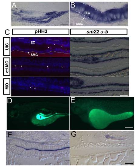
Expression of gata6 in the gut and characterization of differentiation defects in the miR-145 morphants. (A)Alongitudinal section of a 96 hpf wild-type embryo shows strong expression of gata6 in gut epithelial and smooth muscle cells. (B) Enlargement of (A) highlights staining of gata6 in both layers. White dashed lines distinguish the smooth muscle cell layer and epithelial cell layer. (C) Longitudinal sections of phosphohistone H3 (pHH3) staining (red) in the 96 hpf gut. Nuclei were counterstained with DAPI (blue). No significant difference of pHH3 positive cells (white arrowheads) was found in the different experimental groups in either epithelial or smooth muscle layers. The white line in the uninjected embryo highlights the gut epithelium. In the second column, longitudinal sections of sm22α-b in situ hybridization in 96 hpf embryos shows specific staining in the smooth muscle cell layer, and highlights abnormal morphology of both epithelial and smooth muscle cells in miR-145 morphants as compared to controls. (Dand E) The wild-type gut (g) is strongly fluorescent at 120 hpf after DAF-2DA staining (Green) in comparison with miR-145 morphants. (F and G) Strong alkaline phosphatase activity is observed in the gut lumen of wild-type embryos but is absent in miR-145 morphants. SMC, smooth muscle cell (white arrow); EC, epithelial cell; ctl. MO, control morpholino. (Scale bars, A: 50 μm; B: 10 μm; C and G; 25 μm; D: 200 μm.) |

gata6 expression level are not modulated by miR-145 at early embryonic stages. (A and B) gata6 expression levels (white arrowhead) are unchanged in myocardial precursors of uninjected wild-type embryos and miR-145 morphants at 16–19 hpf. (C and D) gata6 expression is significantly increased in miR-145 morphants at 48 hpf. (Scale bars, 200 μm.) (E) There is no significant change in gata6 expression in miR-145 morphants at 16 hpf and 24 hpf by qPCR. EXPRESSION / LABELING:
|
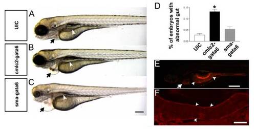
Characterization of tissue-specific gata6 overexpression on development. (A–C) Pericardial edema (black arrow) results from injection of either SMA:Gata6 or CMLC2:Gata6, but only SMA:Gata6-injected embryos show an abnormal gut tube (white arrowhead) at 96 hpf. (D) The percentage of embryos exhibiting the abnormal gut morphology was analyzed by using Student’s t test (P < 0.05). A significantly higher percentage of embryos with abnormal gut development is found in SMA:Gata6 injected embryos. Bars,mean±SEM. *, P<0.05 (n>50 per group). (E) Lateral view of a control Tg (SMA: mCherry) transgenic embryo at 96 hpf. The heart (white arrow) and gut smooth muscle (white arrowheads) show expression of mCherry. (F) Longitudinal section of a 96 hpf Tg (SMA: mCherry) embryo shows that mCherry is specifically expressed in visceral smooth muscle cells (white arrowheads). (Scale bars, C: 200 μm; E: 400 μm; F: 50 μm.) |

Unillustrated author statements PHENOTYPE:
|