- Title
-
The phytoestrogen genistein affects zebrafish development through two different pathways
- Authors
- Sassi-Messai, S., Gibert, Y., Bernard, L., Nishio, S., Ferri Lagneau, K.F., Molina, J., Andersson-Lendahl, M., Benoit, G., Balaguer, P., and Laudet, V.
- Source
- Full text @ PLoS One
|
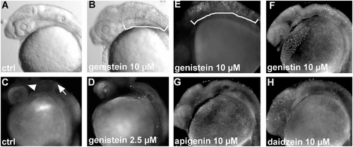
Genistein induces apoptosis during zebrafish development (A) 24 hpf photo of an untreated embryo and an embryo treated with 10 μM of genistein showing necrosis in the posterior hindbrain and in the anterior spinal cord marked by the white bracket (B). (C–H) Apoptotic cell detection at 24 hpf using acridine orange. (C) Control embryo with few apoptotic cells in the midbrain (arrowhead) and the hindbrain (arrow). (D, E) Embryo exposed to 2.5 and 10 μM of genistein showing an increased number of stained cells especially in the hindbrain and a high number of apoptotic cells in the hindbrain and anterior spinal cord when high concentrations of genistein are used (bracket in F). (F–H) Apoptosis mainly found in the hindbrain (arrows) induced by compounds related to genistein: genistin (F), apigenin (G) and daidzein (H) each at 10 μM. Using daidzein, numerous apoptotic cells are detected in the forebrain in comparison to what is observed with the other compounds. |
|
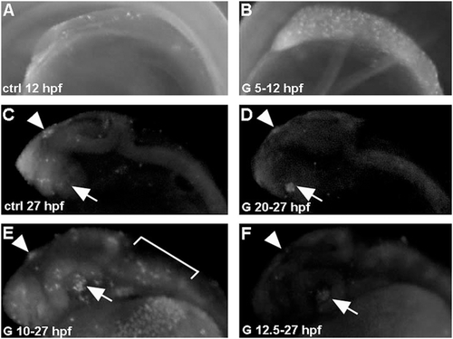
Genistein-induced apoptosis is developmental stage-dependent. Embryos were treated with 2.5 μM genistein for 7 hours starting at 5 hpf (B) or 20 hpf (D) and stained with acridine orange. A high number of apoptotic cells, compare with control (A), are detected in embryos treated from 5 hpf onwards. A similar localization (retina and epiphysis, arrow and arrowhead, respectively) and small number of apoptotic cells are detected in control (C) and genistein-treated embryos from 20 hpf onwards (D). (E) Treatment from 10 hpf onwards induces a strong apoptosis detected at 27 hpf (arrows), while a similar treatment started at 12.5 hpf did not induce apoptosis at 27 hpf (F). |
|
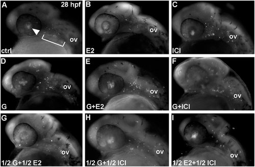
Genistein-induced apoptosis is estrogen receptor independent. (A,B) estradiol does not induce apoptosis. Embryos treated with 1 μM of E2 from 5 hpf onwards display the same pattern of apoptosis (retina and epiphysis) as controls (compare A and B). In contrast embryos treated with 1 μM ICI 182,780 exhibit an increased number of apoptotic cells in the hindbrain (C). Genistein induce an apoptosis at 5 mM as shown Figure 1 (D) and cotreatments with E2 (E) or ICI 182,780 (F) does not modify this effet. Effects of treatments with half dose (0.5 μM for E2 and ICI 182,780 and 2.5 μM for genistein) led to the same conclusions. See Supplementary Figure 2 for the quantitative analysis of these results. |
|
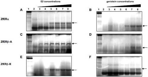
Genistein binds the three zebrafish ERs. Limited proteolysis assays of in vitro translated zebrafish ERα, ERβ-A and ERβ-B with E2 as ligand (A,C,E, respectively) and genistein as ligand (B,D,F, respectively). For each proteolysis panel, the first line represents the undigested protein, line 2 shows digestion of the receptor in the absence of ligand, lines 3 to 8 show digestion of the receptor in the presence of 10 fold increased concentrations of E2 and genistein from 10-8 M to 10-3 M. |
|
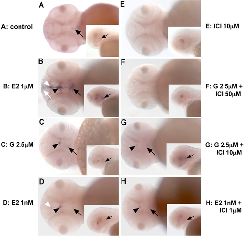
Genistein regulates aromatase-B expression in the brain in an estrogen receptor-dependent manner. In toto in situ hybridization showing the expression of aromatase-B in the zebrafish brain at 48 hpf. (As) Control embryo with weak expression detected in mediobasal hypothalamus (MBH) (arrowheads). (Bs) After genistein exposure, expression is observed in MBH (arrowheads) and in the preoptic area (POA) (arrows). ER antagonists, like ICI, do not induce aromatase-B expression (Cs). The weak staining observed in the MBH in controls is no longer detected in ICI-treated embryos. Cotreatment of genistein and ICI abolishes aromatase-B expression (Ds), but a low dose of ICI does not totally block genistein-induced aromatase-B expression (compare Ds with Es). E2 at high concentrations induces expression of aromatase-B in the MBH (arrowhead), POA (arrow) and in the telencephalon (Tel). When a lower concentration of E2 (1 nM) is used, expression is no longer detected in the anterior brain (Tel) but remains in the MBH (arrowhead) and POA (arrow) as after genistein exposure (compare Gs and Bs). As for genistein, cotreatment of ICI with low concentrations of E2 strongly reduces aromatase-B expression (Hs). |
|
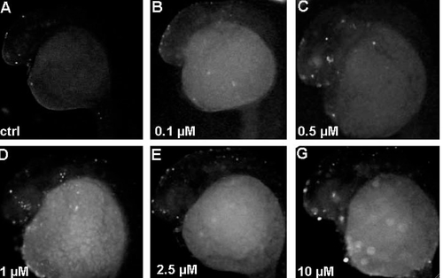
Detection of apoptosis in genistein treated fishes using TUNEL assays, a direct method for the presence of fragmented DNA (Lawry 2004). Fishes were treated at 5 hpf and labeled at 24 hpf with the indicated concentration of genistein. We observe a dose-dependent activation of apoptosis starting at 0.5 μM. |

