
- Title
-
Disruption of AP1S1, causing a novel neurocutaneous syndrome, perturbs development of the skin and spinal cord
- Authors
- Montpetit, A., Côté, S., Brustein, E., Drouin, C.A., Lapointe, L., Boudreau, M., Meloche, C., Drouin, R., Hudson, T.J., Drapeau, P., and Cossette, P.
- Source
- Full text @ PLoS Genet.
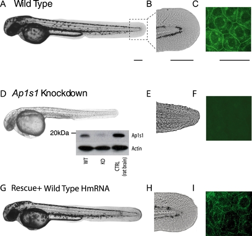
Morphological phenotype of Ap1s1 knockdown zebrafish is rescued by over expression of human AP1S1. Transmitted light images of 48 hpf Ap1s1 KD larvae show their smaller size, reduced pigmentation (D) and skin disorganization (E) compared to the WT (A, B) and the rescued larvae (G,H). Immunofluorescence of wholemount zebrafish using anti-Ap1s1 antibody showing localization of Ap1s1 to the plasma membrane (C, polygonal) and to a well defined perinuclear ring, in both normal (C) and rescued larvae (G), whereas only a residual and diffuse staining could be observed in KD larvae (F). Western blot analysis (D, inset) indicates nearly complete knockdown of Ap1s1 protein (WT = wild-type, KD = knockdown, CTRL = control rat brain proteins). To normalize the western blot analysis, proteins extracted from WT, KD and CTRL larvae were incubated with anti-actin. Scale bars in (A, D, G) = 100 μm, (B, C, E, F, H, I-Ciii) = 50 μm. |
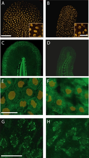
Ap1s1 knockdown is associated with abnormal distribution of laminin and cadherin. Despite changes in the size and shape of the tail, p63-labelled nuclei (orange) of basal keratinocytes were present in KD larvae. (B). The insets show enlarged views of small groups of keratinocytes. Immunolabelling for laminin (green) in WT (C) showed normal distribution of the basement membrane along the fin fold margin, whereas in the KD larvae the residual laminin labeling was diffuse and disorganized (D). Compared to WT keratinocytes (E) labeled with p63 (orange), cadherin (green) is decreased at the cell membrane of the KD larvae (F). However, the localization of cytokeratin in WT (G) and in KD larvae (H) appeared similar. Scale bars: 10 μm, Insets in A, B, = 20 μm. |

Abnormal behavioral phenotype and impaired development of spinal neural network of Ap1s1 knockdown zebrafish. Consecutive images from films illustrating the response to touch in 48 hpf wild type (WT) (A) and Ap1s1 KD larvae (F). The WT larva reacted to touch by swimming away. In contrast, the Ap1s1 KD larvae exhibit slow and impaired reaction. Immunostaining in wholemount 48 hpf WT (B–E) and Ap1s1 KD larvae (G–J) illustrating the reduced axonal labeling (anti-acetylated tubulin; B and G red, the arrows point to the ventral roots), the reduced number of newly born cells (anti-HU antibody; red in C and H), and the number of interneurons (anti-Pax2 antibody; E and J green) in the Ap1s1 KD larvae. In turn, the number of motoneurons (anti-HB9 antibody; D and I, red) was similar both in WT and Ap1s1 KD. Scale bars: 25 μM. |
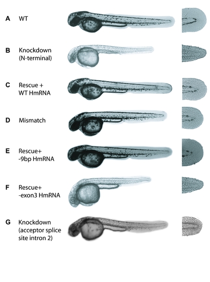
Representative transmitted light images of 48 hours-old zebrafish larvae illustrating the phenotype under different experimental conditions. (A) Wild type (WT), non injected larvae. (B) Knockdown larva, injected with AMO targeting the translation start site of Ap1s1 (N-terminal). (C) Larva rescued by co-injection of AMO and human WT mRNA (HmRNA). (D) Larva injected with a mismatch AMO. (E) Larva rescued by co-injection of AMO and -9bp Human mRNA. (F) larva co-injected with AMO and -exon3 HmRNA. (G) Knockdown larva, injected with AMO targeting the acceptor splice site (intron 2) of Ap1s1. PHENOTYPE:
|