- Title
-
Germ line control of female sex determination in zebrafish
- Authors
- Siegfried, K.R., and Nüsslein-Volhard, C.
- Source
- Full text @ Dev. Biol.
|
Zebrafish lacking germ cells make testes. H&E stainings of adult (7 month old) wild-type and germ line deficient testes. (A) Wild-type testis. The testis is organized in tubules surrounded by basement membrane, outlined in black (A′). Within the tubules, cysts of germ cells undergo spermatogenesis synchronously, outlined in blue (A′). Spermatozoa (sz) are released into the center, which connects to the efferent ducts. Sertoli cells (arrowheads and inset) are found within the tubules and Leydig cells (L) are in the interstitial spaces, colored purple (A′). (B) Germ line deficient testis. Testis tubules are formed, outlined in black (B′). Sertoli cells are lining the inside of the tubules (some marked by arrowheads and shown in inset). Leydig cells (L) are intertubular, colored purple (B′). Vasculature is also present in intertubular regions. sz, spermatozoa; st, spermatids; sg, spermatogonia. Scale bars are 10 μm. PHENOTYPE:
|
|
Adult germ line deficient gonads exhibit expression of genes normally expressed in wild-type testes. (A) RT-PCR of genes normally expressed in the ovary and/or testis. Each experiment was done on two independent samples. Negative controls lacking reverse transcriptase are in alternating lanes (c). (B-C) Detection of 3βHSD activity in Leydig cells using a chromatographic assay. (B) Wild-type and (C) germ line deficient testes have 3βHSD activity in intertubular regions where Leydig cells reside. Scale bars are 10 μm. EXPRESSION / LABELING:
|
|
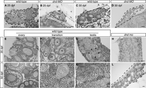
Histological analysis of wild-type and germ line deficient gonads during sex-differentiation. Hematoxylin and Eosin stained sections of gonads from 25 dpf to 50 dpf. The gonad is outlined by a hashed line in some panels. (A-D) Gonads from wild-type and germ line deficient fish at 25 and 30 dpf. Wild-type gonads have stage 1b perinuclear oocytes with associated somatic pre-follicle cells (white arrowheads) whereas dnd-MO treated siblings have only somatic gonadal tissue. (E-G) Wild-type gonads at 35 dpf. Three developmental phases are found: (E) juvenile ovary, where healthy stage 1b oocytes are present; (F) “transition”, where the oocytes are degenerating (*) and the gonad is in the process of transitioning to a testis; and (G) testis, where oocytes are no longer present, but spermatogonial cells are present in cyst-like structures (black arrowheads denote one spermatogonial cyst). (H) Gonads from 35 dpf dnd-MO injected fish have no germ cells and no apparent organization of testes structures. (I-K) Wild-type gonads at 50 dpf, individuals are at various developmental stages: (I) oogenesis progresses in some animals; (J) gonads in transition are still found, where oocytes are intermixed with testis tissue; and (K) progression of spermatogenesis and clear organization of testis tubules is also apparent. (L) At 50 dpf, morphogenesis of testes tubules becomes apparent in germ line deficient testes. Scale bars are 10 μm. |
|
In situ hybridization on gonads during sex-differentiation. (A-H) At 25 and 30 dpf gonads from nearly all fish exhibit expression of sox9a and cyp19a1a. (I-N) At 35 dpf dimorphic gene expression is apparent. In wild-type, dimorphic expression of sox9a is apparent (I,M) and expression is strong in all gonads from dnd-MO injected fish (J). cyp19a1a expression also becomes dimorphic in wild-type (K,N) and is no longer detectable in gonads from dnd-MO injected fish. The sox9a transcript is generally diffuse in appearance and, at 25 and 35 dpf, is primarily localized at the borders of the organ in wild-type whereas cyp19a1a transcript is localized in distinct spots presumably small somatic cells. In panels where extra-gonadal tissue is present, the gonad is denoted with black arrowheads. White arrowheads point to pigment cells. |
Reprinted from Developmental Biology, 324(2), Siegfried, K.R., and Nüsslein-Volhard, C., Germ line control of female sex determination in zebrafish, 277-287, Copyright (2008) with permission from Elsevier. Full text @ Dev. Biol.


