- Title
-
Enhancer detection in zebrafish permits the identification of neuronal subtypes that express Hox4 paralogs
- Authors
- Punnamoottil, B., Kikuta, H., Pezeron, G., Erceg, J., Becker, T.S., and Rinkwitz, S.
- Source
- Full text @ Dev. Dyn.
|
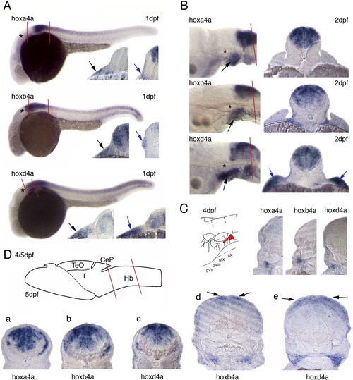
Hoxa4a, hoxb4a, and hoxd4a expression in developing embryos and larvae. A: In situ hybridized embryos at 1 dpf. The otic vesicle is marked by an asterisk. Transverse sections were performed through the anterior r7 and posterior. The levels of sections are indicated. Black arrows indicate signal in the epibranchial placode. Hybridization signals below the placode indicate expression in the branchial arches. Blue arrows in further posterior sections point to (1) signal adjacent to the somitic mesoderm (hoxa4a), (2) signal in lateral line neuromasts (hoxb4a), and (3) expression in the fin anlage (hoxd4a). B: Hox4 expression in the posterior hindbrain and in the branchial arches (black arrows) at 2 dpf with 20μm sections on 3rd somite level. The blue arrows point to signals in the fin (hoxd4a). C: Schematic drawing of the ganglia VII to X and 20μm sections performed through hybridized larvae at 4dpf with mRNA-transcript in the vagal ganglion. D: Schematic drawing of a 5-dpf zebrafish brain modified from Mueller and Wullimann ([2005]) indicating the level of sections through the hindbrain (a-c) and through the cerebellum (d,e). The black arrows point to hybridization signal in the cerebellum. CEP, cerebellar plate; Hb, hindbrain; TeO, tectum opticum; T, midbrain tegmentum. |
|
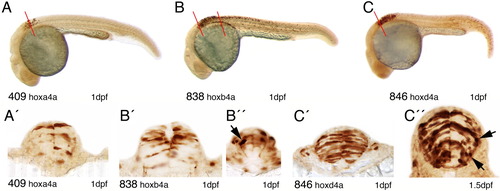
YFP expression in 1-dpf embryos of hoxa4a, hoxb4a, and hoxd4a enhancer detection lines as detected by immunohistochemistry. A-C: YFP expression pattern in the hindbrain and spinal cord of the transgenic lines CLGY409, CLGY838 and CLGY846. A′: Hoxa4a-YFP expression in elongated neuroepithelial cells in r8. The level of section is indicated in A. B′, B″: Transverse sections through the hindbrain of CLGY 838 show hoxb4a-YFP expression in elongated neuronal precursor cells on the level of r7 (B′) and r8, which also shows a neuron with a ventromedial projection (B″, black arrow). The levels of sections are indicated in B. C′: Many neuroepithelial cells were YFP positive in r7 of the hoxd4a enhancer detection embryos. The level of section is indicated in C. C″: Section through r7 of hoxd4a embryos at 1.5 dpf, showing YFP-positive postmitotic neurons, with young immature neurons sending out small primary projections (black arrows). EXPRESSION / LABELING:
|
|
Immunostained embryos/larvae of hoxa4a (A), hoxb4a (B, C), and hoxd4a (D) enhancer detection lines with transverse sections on the level of the first (r7) and third somite (r8). A: hoxa4a-regulated YFP expression at 2 dpf in the hindbrain r7 and r8, in the vagal nerve (white arrows), the pectoral fin nerve (yellow arrows), and in some neural crest cells (black arrow). Stained cells are located further ventrally and medially than in the hoxb4a enhancer trap line. B: hoxb4a-YFP expression in embryos at 2 dpf in the posterior hindbrain, in the vagal nerve, vagal ganglion (white arrows), in the pectoral fin nerve (yellow arrows), and some neural crest cells (black arrow). In r8, at the level of the fin motoneuron, there were no YFP-positive neurons in the medial hindbrain, which was also observed at later developmental stages. C: At 3 dpf, hoxb4a-regulated YFP was detected in ventrally located fiber tracts that grew from the posterior hindbrain towards the cerebellum (blue arrow). The white arrows point to the vagal and the yellow arrows to the pectoral fin nerve. Neural crest are marked by the black arrow. D: Hoxd4a-YFP expression at 3 dpf in the posterior hindbrain, in the vagal nerve and the vagal ganglion (white arrows), in the heart wall and the fin (black arrows). In contrast to the hoxb4a enhancer detection line, cells are also stained in the medioventral hindbrain. |
|
Sections through anti-GFP (islet1 GFP transgenic line) and anti-YFP (hoxb4a and hoxd4a enhancer detection lines) immunostained larvae. A: Islet 1 expression at 7 dpf in the vagal nucleus (dorsal black arrows) and the vagal visceromotor nerve (blue arrows). B: Hoxb4a regulated YFP expression in a broad hindbrain domain and in a subpopulation of the vagal nerve (blue arrows). The black arrows point to neuropithelial cells, which are YFP positive. C-E: Hoxd4a-YFP expression in 5-dpf larvae. C: A section anterior of the first somite. The blue arrows point to weakly stained fibers of the vagal nerve exiting the hindbrain. Vagal ganglion cells and a fine fiber innervating the pharynx (green arrows) are also YFP positive. D: One section further posterior to C with YFP-labeled vagal ganglion cells that project towards the pharynx (green arrows). E: A magnification of C with focus on the fine fibers that innervate the pharynx. Arrows follow the same color code as in C and D. Ph, pharynx; pi, pigment. |
|
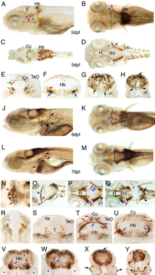
Anti-YFP immunostainings of hoxb4a enhancer detection larvae at 5 dpf (A-H), 6 dpf (J, K), and 7 dpf (L-Y). The arrows have the following color code. Red, blue: 2 precerebellar fiber tracts; orange: cells in ventral tegmentum; green: cells in cerebellum; light blue: fiber tract projection towards tectum. The yellow and black arrows are explained below. A,B: Whole-mount larvae at 5 dpf. The yellow arrow marks a nerve innervating the fin. C,D: Horizontal sections through a 5-dpf larva on level of the dorsal hindbrain/cerebellum (C) and ventral hindbrain (D). E-H: Transverse sections through a 5-dpf larva. The black line frames a tangential stripe pattern of YFP-positive neurons. The black arrows point to neurons in the ventral hindbrain and to a neuroepithelial cell located dorsally. J,K: YFP distribution in 6-dpf larvae. The left yellow arrow points to the vagal nerve, the right yellow arrow marks a nerve innervating the fin. Black arrows point to neural crest cells. L,M: Anti-YFP-stained larvae at 7 dpf. Black arrows mark neural crest cells. N-Q: Horizontal sections through 7-dpf larvae, in a row from dorsal to ventral. The black arrows mark cells around the somites. R-Y: Transverse sections through a 7-dpf larva, in a row from anterior to posterior. The lateral and ventral black arrows point to cells around the somites and the dorsal black arrow to a single stained neuroepithelial cell. The asterisk marks the inner ear. D, diencephalon; Cc, corpus cerebelli; H, hypothalamus; Hb, hindbrain; Rv, rhombencephalic vesicle; s1, first somite; T, midbrain tegmentum; TeO, tectum opticum; Va, Valvula cerebelli. |
|
Anti-YFP staining of the hoxd4a enhancer detection line, at 4 dpf (A-D), 5 dpf (E, F), 6 dpf (G-L). The arrows have the following color code. Red, blue: 2 different precerebellar fiber tracts; orange: cells in ventral tegmentum; green: cells in cerebellum; yellow: vagal nerve and vagal ganglion. The black and grey arrows are explained in the text. A: YFP distribution in the posterior hindbrain. The black arrow marks cells in the anterior hindbrain. B-D: Transverse sections through a 4-dpf larva from anterior to posterior. The black arrows indicate neuroepithelial cells and the grey arrows two symmetrical clusters of neurons. The hindbrain neurons followed tangential stripes (framed by a black line). E,F: Horizontal sections through a 5-dpf larva on the level of the dorsal (E) and ventral (F) hindbrain. Black arrows point to YFP-positive cells in the hindbrain. G,H: Whole mount stained larva at 6 dpf with YFP expression in the posterior hindbrain. J-L: Transverse sections through a 6-dpf larva in a row from anterior to posterior. Black arrows mark contralaterally projecting hindbrain neurons. The asterisk marks the inner ear. Cc, corpus cerebelli; H, hypothalamus; Hb, hindbrain; s, somite; T, midbrain tegmentum; TeO, tectum opticum. |
|
The reticulospinal formation was labeled in CLGY838 larvae at 4 dpf and cryosections through these specimen were double immunostained for Biotin and YFP. YFP-positive cells are shown in green, the rhodamin-dextan labeled reticulospinal neurons are red. Hoxb4a-YFP cells did not overlap with reticulospinal neurons. A: Transverse section at first somite level. B: Sagittal section through the medial hindbrain. The asterisk marks a region where YFP-positive neurons are absent. C: Magnification of inset from sagittal section. |


