
- Title
-
Nr4a2 is essential for the differentiation of dopaminergic neurons during zebrafish embryogenesis
- Authors
- Luo, G.R., Chen, Y., Li, X.P., Liu, T.X., and Le, W.D.
- Source
- Full text @ Mol. Cell Neurosci.

Distribution of nr4a2b transcripts throughout the early developmental stages of zebrafish embryos. Anterior is to the left and dorsal is up. (A–D) nr4a2b is a maternal expressed gene and is universally expressed from 0.75 hpf to 12 hpf stage. (E) The first specific transcripts are found in brain at about the stage of 18 hpf (white arrows). (F) Two groups of nr4a2b+ cells appear at 24 hpf. One is near OB in tel (black heads), and the other is around the rh (white arrowheads). A group of nr4a2b+ cells emerges in PT (white arrows). (G–J) The nr4a2b+ cells in PT area (white arrows) and in tg (black arrows in the lateral view) are expanded from 30 hpf to 120 hpf. At the stage of 48–120 hpf, nr4a2b transcripts display a complex pattern (I and J). It is expressed at MO (black arrows in the dorsal review) and ret (black arrowheads). MO, medulla oblongata; OB, olfactory bulb; PT, posterior tuberculum; rh, rhombencephalon; ret, retina; tel, telencephalon; tg, tegmentum. |
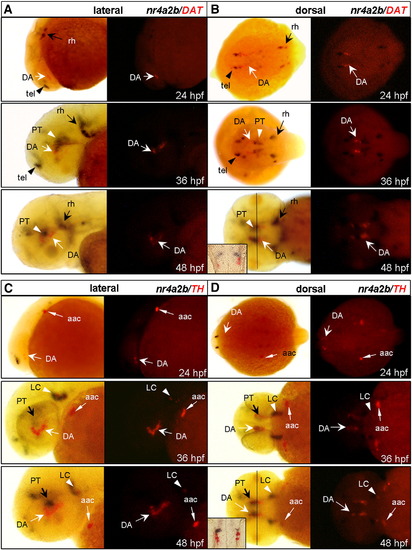
nr4a2b (black) has little co-expression with TH and DAT (red) in the PT area. Anterior is to the left and dorsal is up. (A and B) Lateral and dorsal views show the nr4a2b and DAT expression patterns of 24, 36, 48 hpf embryos. DA neurons (white arrows) are located in the PT area, while nr4a2b+ cells are expressed at the dorsal (white arrowheads). (C and D) Lateral and dorsal views show the nr4a2b and TH expression pattern of 24, 36, 48 hpf embryos. Most of the TH+ neurons (white arrows) surround by nr4a2b+ cells (black arrows) in basal forebrain and some of them are closely located with nr4a2b+ cells in LC (white arrowheads). Solid lines in B and D correspond to transverse section presented as the insets. Dorsal is up. Fast Red labeling of TH+ or DAT+ cells (RITC filter set) is shown at the right panel. aac, arch-associated catecholamine cells; DA, dopamine neuron; DAT, dopamine transporter; LC, locus coeruleus; PT, posterior tuberculum; rh, rhombencephalon; tel, telencephalon; TH, tyrosine hydroxylase. |

nr4a2b (red) is expressed in the neurog1+ progenitor (black) in the early embryogenesis. (A and B) Co-expression of neurog1 and nr4a2b in basal forebrain of 24 hpf embryos (white arrowheads). (C and D) Co-expression of neurog1 and nr4a2b in basal forebrain of 36 hpf embryos (white arrows). (E and F) Two sagittal sections of the PT area are marked by white rectangular box in panel C and two transverse sections are marked by solid white line in panel D, while black arrows indicate the single cell co-expressing neurog1 and nr4a2b. (G and H) Expression of neurog1 and nr4a2b in basal forebrain of 48 hpf embryos. Anterior is to the left and dorsal is up. Lateral view is in the left panel and dorsal view is in the right panel. Fast Red labeling of nr4a2b+ cells (RITC filter set) is shown at the right panel. |

Nr4a2 is essential during the development of DA neurons in the PT area. (A) Western blot analysis of 48 hpf embryo lysates with Nr4a2 monoclonal antibody (upper panel), as well as β-actin monoclonal antibody as a loading control (lower panel). Protein lysates from 10 embryos are loaded in each lane. (B) Western blot analysis of Nr4a2 and TH level of the 5-mm injected, Nr4a2-deficient and rescued embryo lysates at 48 hpf; β-actin monoclonal antibody is used as a loading control. (C) In the Nr4a2a-, Nr4a2b- and Nr4a2-deficient embryos, TH staining is significantly decreased not only in Pr, PO and OB (black arrows), but also in the PT area (white arrows). Anterior is to the left. (D) Percentage of Nr4a2a-, Nr4a2b- and Nr4a2-deficient embryos that have the significantly decreased TH expression in Pr, PO, OB and the PT area. (E) Exogenous mouse Nr4a2 transcripts can partially rescue the loss-of-function of endogenous Nr4a2. Fewer TH+ and DAT+ neurons in the PT area are shown in nr4a2mo-injected embryos at 48 hpf and the TH+ and DAT+ neurons in the PT area can be partially rescued after injection with mouse Nr4a2 mRNA (white arrows), with the TH+ neurons in the LC area as control (black arrowheads). (F) Bar scale of the HPLC analysis for DA content. The level of DA is decreased by 79.5% (p = 0.0027) in the embryo after nr4a2mo treatment and it is recovered by 55.5% (p = 0.0029) after mouse Nr4a2 mRNA injection. DAT, dopamine transporter; LC, locus coeruleus; OB, olfactory bulb; PO, preoptic area; Pr, pretectum area; TH, tyrosine hydroxylase. |

Knockdown of Nr4a2 results in the increase of nr4a2b+ cells and the inhibition of DA neuron differentiation. (A and B) In the embryos injected with nr4a2mo, the number of nr4a2b+ cells (black) is increased while the number of TH+ neurons is decreased (red) (white arrows). (C) Bar scale to show that the number of nr4a2b+ cells (red) in nr4a2mo is increased by 57.5% (p = 0.0172) compared to 5-mm injected embryos. (D) BrdU incorporation and TUNEL assay to show that there is no obvious cell proliferation or apoptosis in the PT area. White rectangles show the position of the PT area between two eyeballs as the dashed lines show. |

RT-PCR to show the expression level of nr4a2b transcripts throughout the developmental stages from 0.75 to 120 hpf with gapdh as loading control and negative control. |

RT-PCR to show the abnormal splicing products of nr4a2a and nr4a2b in nr4a2amo and nr4a2bmo-injected embryos at 48 hpf, respectively, with 5-mm and nr4a2mo-injected embryos as control. The gapdh was used as a loading control. Arrows and arrowheads indicate the normal and abnormal splicing products, respectively. EXPRESSION / LABELING:
|
Reprinted from Molecular and cellular neurosciences, 39(2), Luo, G.R., Chen, Y., Li, X.P., Liu, T.X., and Le, W.D., Nr4a2 is essential for the differentiation of dopaminergic neurons during zebrafish embryogenesis, 202-210, Copyright (2008) with permission from Elsevier. Full text @ Mol. Cell Neurosci.