
- Title
-
psoriasis regulates epidermal development in zebrafish
- Authors
- Webb, A.E., Driever, W., and Kimelman, D.
- Source
- Full text @ Dev. Dyn.
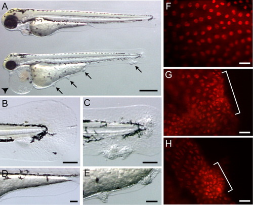
psoriasis mutants have epidermal defects. A: Wild-type and psoriasis (bottom) embryos at 60 hours postfertilization (hpf). psoriasis embryos develop pericardial edema (arrowhead) and epidermal aggregates (arrows). B-E: Wild-type (B,D) and psoriasis (C, E) embryos at 60 hpf showing regions of ectopic skin aggregates in psoriasis. F-H: Wild-type (F) and psoriasis (G,H) embryos stained for the epidermal-specific nuclear marker, p63. Note the presence of p63 within epidermal clumps (brackets). Anterior is to the left in A-E and to the upper left in F-H. Scale bars = 250 μm in A, 50 μm in B-E, 12.5 μm in F-H. |
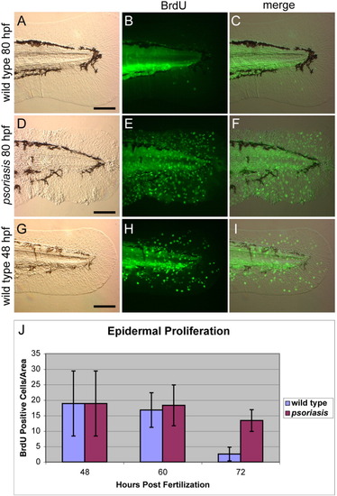
psoriasis embryos fail to cease epidermal proliferation at 80 hours postfertilization (hpf). A-C: The epidermis of wild-type embryos is 5′-bromodeoxyuridine (BrdU) -negative (B) at 80 hpf, indicating cessation of epidermal proliferation late in embryogenesis. D-F: Epidermal cells in psoriasis mutants are BrdU-positive at 80 hpf (E). G-I: The level of proliferation observed in psoriasis embryos is similar to that observed in 48 hr wild-type embryos (H). Anterior is to the left. Brightfield images are shown in A, D, and G, BrdU-positive cells are green in B, E, and H. C, F, and I are merged brightfield and fluorescent images. J: Quantification of cell proliferation at 48, 60, and 72 hpf. Scale bars = 50 μm. PHENOTYPE:
|
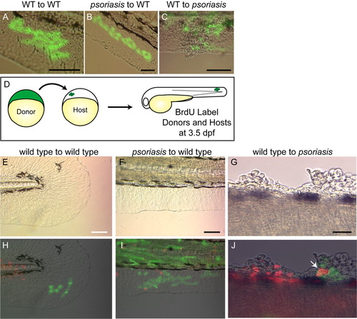
psoriasis acts non-cell-autonomously. A: Control transplant showing wild-type cells transplanted into a wild-type host epidermis. B: psoriasis mutant cells transplanted into a wild-type host contribute to epidermis and display wild-type morphology. C: Wild-type cells transplanted into a psoriasis mutant contribute to epidermal aggregates. D: Overview of transplantation experiments combined with 5′-bromodeoxyuridine (BrdU) assays. Cells from fluorescein-labeled donors were transplanted to unlabeled hosts at 4 hours postfertilization (hpf). At 3.5 days postfertilization (dpf), cell proliferation was assayed by BrdU-labeling donors and hosts. E-G: Brightfield images of host embryos. H-J: Merged images showing transplanted cells (green) and BrdU-positive cells (red) are shown below the corresponding brightfield images. E,H: In wild-type to wild-type transplants, transplanted epidermal cells are BrdU-negative, showing that the wild-type embryo and transplanted cells have halted proliferation. F,I: psoriasis cells transplanted into a wild-type host are BrdU-negative. G,J: A psoriasis host containing a BrdU-positive wild-type epidermal cell (arrow) at 80 hpf. Anterior is to the left; all embryos shown at 80 hpf. Scale bars = 100 μm. |
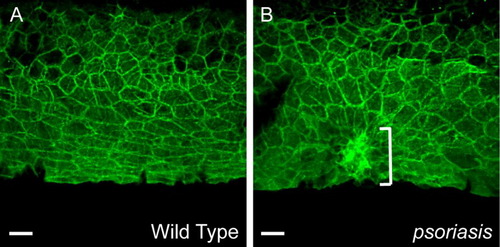
psoriasis mutants maintain epidermal contacts and normal cell morphology away from epidermal aggregates. A: Wild-type epidermal cells localize cadherins to cell membranes. B: psoriasis mutants display normal cadherin localization in regions away from epidermal aggregates. Cells within epidermal clumps express cadherins (bracket), but lose flattened wild-type morphology. Scale bars = 12.5 μm. PHENOTYPE:
|
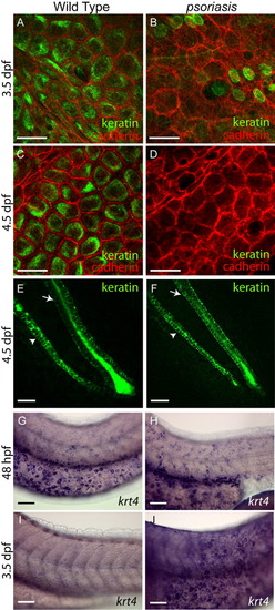
psoriasis mutants fail to express differentiation-specific keratins. A-D: Keratin expression in epidermal cells. Wild-type epidermal cells express and localize keratins basally at 3.5 (A) and 4.5 (C) days postfertilization (dpf), whereas psoriasis mutant epidermal cells have greatly decreased keratin expression at 3.5 (B) and 4.5 (D) dpf. The round green cells in B are unidentified, but are likely to be neuromast precursors based on regular spacing along the anterior-posterior axis in the position of the future lateral line. Cadherin staining (red) marks epidermal cell membranes. E,F: Keratin expression along the axis of the tail. Arrowheads indicate hypochord, and arrows indicate neural tube, respectively. Keratin expression is normal in this region of wild-type (E) and psoriasis mutants (F). E and F are lateral views with anterior to the upper left. G-J: In situ hybridizations for krt4. Wild-type embryos express krt4 at 48 hours postfertilization (hpf; G) and at lower levels at 3.5 dpf (I). psoriasis mutants express krt4 at 48 hpf similar to wild-type embryos (H), but exhibit increased krt4 expression at 3.5 dpf (J). Embryos in all these panels were stained for the same length of time. Anterior is to the left and dorsal is up in G-J. Scale bars = 25 μm in E,F, 50 μm in G-J. PHENOTYPE:
|

Epidermal aggregates contain dead and dying cells. A-C: The epidermis of wild-type embryos is acridine orange-negative. D-F: psoriasis mutants stain with acridine orange only in regions of epidermal cell accumulation. Images are shown as brightfield (A,D), acridine orange (B,E), and merged (C,F). Anterior is to the left and dorsal to the top. Scale bars = 50 μm. PHENOTYPE:
|

Unillustrated author statements PHENOTYPE:
|