
- Title
-
Complex cell rearrangements during intersegmental vessel sprouting and vessel fusion in the zebrafish embryo
- Authors
- Blum, Y., Belting, H.G., Ellertsdottir, E., Herwig, L., Lüders, F., and Affolter, M.
- Source
- Full text @ Dev. Biol.

Complex distribution of junctional proteins in ISV. (A) Cellular model of ISVs according to Childs et al. (2002). (B) Putative distribution of cell junctions (red rings) in an ISV in which endothelial cells are arranged in a head-to-tail fashion forming a seamless tube. (C–E) Confocal projection of 36 hpf fli1:EGFP (red) embryos labeled with an anti-ZO-1 antibody (green). ZO-1 protein is mostly detected as two “stripes” (arrows in panels C and D) along the stalk of the ISV, and also in parts of the DLAV. Sometimes, ZO-1 is absent from short regions of the ISVs, including the DLAV (E, arrowheads). (F–I) ZO-1 outlines single cells. 30 hpf wild-type fish injected with flk1:RAS-GFP (red) plasmid and labeled with anti ZO-1 antibodies (green). (F and G) Putative single stalk cells are entirely lined by ZO-1. (H and I) Putative tip cells show ring-like ZO-1 pattern at their base, where they make contact with stalk cells (arrows). Abbreviations: da: dorsal aorta, nc: notochord, sb: somite boundary. Scalebars: 20 μm. EXPRESSION / LABELING:
|

Endothelial cells are paired in the ISV. (A–D) Confocal projection of single cells expressing mCherry under control of the flk1 promoter (shown in green for better contrast) in fli1:EGFP transgenic embryos (red) in ISVs at 36 hpf (A–C) and 48 hpf (D) of development. (B) T-shaped cell embedded in the DLAV, (C, D) extended stalk cells. In all cases, mCherry-expressing cells appear to make up only part of the circumference of the tube (arrows), and appear paired or aligned with cells expressing EGFP only. (E) Single section (X–Y plane) of the cell shown in panel D. (F) Single cross section (Y–Z plane) of the ISV showing the lumen and the surrounding cells. (G, H) Single cross sections of ISVs at 48 hpf showing ZO-1 staining (arrows) between cells surrounding the lumen (2 cells in panel G, 3 cells in panel H). (G′, H′) Schematic representation of the cellular arrangement shown in panels G and H. Scalebars: 20 μm. |
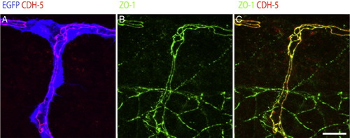
CDH5 colocalizes with ZO-1 in endothelial cells. (A–C) Confocal projections of 36 hpf fli1:EGFP (blue) transgenic embryos labelled with anti CDH5 (red) and anti ZO-1 (green) antibodies. (A) CDH5 and the GFP show that CDH5 labels exclusively junctions of endothelial cells. (B) ZO-1 labels endothelial cells as well as cells in the notochord and the neural tube. (C) CDH5 and ZO-1 show co-localization (yellow) of the two junctional proteins in endothelial cells. Scalebars: 20 μm. EXPRESSION / LABELING:
|
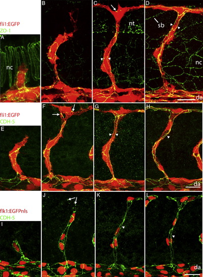
Dynamic expression of junctional proteins during ISV formation. Confocal projections of fli1:EGFP embryos labelled with anti-ZO-1 (A–D) or CDH5 (E–H) antibodies and of flk1:EGFPnls embryos labelled with CDH5 (I–L) antibody at different developmental stages. (A, E, I) 22–24 hpf, (B, F, J) 26–28 hpf, (C, G, K) 30 hpf, (D, H, L) 36 hpf. Already at early stages (A, E) both proteins are localized along the stalk of the ISVs, presumably between putative tip cells on the one hand, and between stalk cells on the other hand. Spots and lines (arrows in panels C, F and J) of ZO-1 and CDH5 are visible when tip cells start to extend in anterior and posterior direction to eventually form the DLAV. Arrowheads point to parallel junctions running along the axis of the ISV. Abbreviations: see Fig. 1, nt: neural tube. Scalebars: 20 μm. EXPRESSION / LABELING:
|

Endothelial cell behaviour during formation of the DLAV. The figure depicts different ISVs within a single embryo. (A–C) ZO-1 pattern during vessel fusion. Confocal projections of 30 hpf fli1:EGFP fish (red) labeled with anti ZO-1 (green). (A) ZO-1 accumulates at the contact site of two putative tip cells, which then crawl over each other resulting in a small (B) oval-shaped ZO-1 pattern, which eventually expands to an extended oval (C). (A′–C′) Schematic representation of the ZO-1 pattern shown in panels A–C. (D, E) Picture series of supplementary movies 1 and 2 showing dynamic cell behaviour during ISV formation. Movie (D) shows a tip cell expressing mCherry in a flk1:EGFP embryo. Within 5 h, the tip cell migrates dorsally and expands in anterior and posterior direction to fuse and form the DLAV. After first contact (D″) tip cells “crawl” over each other to some extent (arrow in panel D′″). Movie (E) shows a single stalk cell migrating dorsally to contribute to the DLAV (arrow in panel E′″). Scalebars: (A–C) 20 μm; (D–D′″) 50 μm; (E–E′″) 100 μm. |

Non-stereotyped behaviour of nuclei in ISVs. Picture series taken from supplementary movies 3 (A) and 4 (B) showing sprouting ISV in flk1:EGFP-NLS embryos. (A) A stalk cell (arrow) divides prior to the tip cell (arrowhead). Outline of the cell can be visualized during cell division. (B) A tip cell nucleus ends up in an adjacent ISV (arrows are following the nuclei). Scalebars: 20 μm. |
Reprinted from Developmental Biology, 316(2), Blum, Y., Belting, H.G., Ellertsdottir, E., Herwig, L., Lüders, F., and Affolter, M., Complex cell rearrangements during intersegmental vessel sprouting and vessel fusion in the zebrafish embryo, 312-322, Copyright (2008) with permission from Elsevier. Full text @ Dev. Biol.