
- Title
-
The netrin receptor UNC5B promotes angiogenesis in specific vascular beds
- Authors
- Navankasattusas, S., Whitehead, K.J., Suli, A., Sorensen, L.K., Lim, A.H., Zhao, J., Park, K.W., Wythe, J.D., Thomas, K.R., Chien, C.B., and Li, D.Y.
- Source
- Full text @ Development
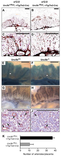
Vascular ablation of Unc5b impacts labyrinthine arterioles. Embryos were harvested at E12.0 from Unc5bflox/flox x Unc5b+/-; Tg(Tie2-Cre)/Tg(Tie2-Cre) (A-D), Unc5b+/-;R26R [Rosa26 Reporter (Soriano, 1999)] x Unc5b+/-; Tg(Tie2-Cre)/Tg(Tie2-Cre) (E,F) or Unc5b+/- x Unc5b+/- (G-J) matings. Placentas were dissected, formalin fixed, hemisected and stained for: smooth muscle actin (anti-smooth muscle actin antibody followed by HRP-conjugated secondary antibody) to identify smooth muscle cells surrounding the arterial bed (A-D); for β-galactosidase activity (X-gal) to identify endothelial expression of Tie2-Cre (E,F); and for Pecam l (anti-Pecam1 antibody followed by HRP-conjugated secondary antibody) to highlight the endothelium (G,H). Ta;-galactosidase activity (X-gal) to identify endothelial expression of Tie2-Cre (E,F); and for Pecam l (anti-Pecam1here is a decrease in the number of robust vertical vessel stalks in placentas lacking UNC5B (B,D,F,H) when compared with their littermate controls (A,C,E,G). This is also shown in I,J, which represent 10 μm cross-sections through samples stained for smooth muscle actin. Arterioles are indicated by arrows; scale bars: 0.5 mm. (K) The number of arterioles per placenta was determined by serial sectioning (10 μm) of hemisected placentas, staining for smooth muscle actin and visually counting all vessels. Paired littermates (three pairs) representing each of the two genotypes were used for all measurements. Error bars are standard deviation. |
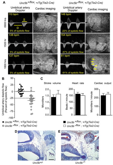
Physiological assessment of embryos. (A) Doppler flow analysis of umbilical arteries and hearts in utero. For longitudinal follow-up of umbilical artery Doppler flow, Unc5b+/flox; +/Tg(Tie2-Cre) and Unc5b-/flox;+/Tg(Tie2-Cre) embryos were monitored on E12 at the time intervals indicated on the left. The ratio of diastolic (D) flow (time velocity integral) over systolic (S) flow is shown as a percentage with a negative number indicative of reverse flow. The ratio decreased progressively over time in the mutant embryo and preceded pericardial effusion (arrows) and bradycardia. (B) Quantitation of umbilical artery diastolic flow reversal. The ratio of umbilical diastolic flow (time velocity integral) over systolic flow was used to quantitate the degree of end diastolic reverse flow and the ratio of -25% of systolic flow (broken line) was chosen as a threshold for significant reversal; 24 Unc5b+/flox; +/Tg(Tie2-Cre) and 23 Unc5b-/flox;+/Tg(Tie2-Cre) embryos were sampled at a single time point at E12.5. (C) The stroke volume, heart rate and cardiac output were recorded at the onset of significant diastolic reverse flow; data from four Unc5b+/flox; +/Tg(Tie2-Cre) and three Unc5b-/flox;+/Tg(Tie2-Cre) are reported; error bars are standard deviation. (D,E) Increased hypoxia in Unc5b-/-embryos. Pregnant females were injected with pimonidazole 2 hours prior to sacrifice and removal of embryos. Sections (10 μm) from the hindbrain region of E12.5 Unc5b+/- (D) and Unc5b-/- (E) embryos were probed with antibodies directed against pimonidazole and HRP-conjugated secondary antibodies, stained for HRP activity (brown, indicated by arrows) and counterstained with hematoxylin (blue). V, trigeminal ganglion. Scale bar: 100 μm. |

Unc5b/Netrin signaling in the placenta. (A,B) Unc5b mRNA localization in the labyrinth. Placentas from E12.5 embryos were hemisected and probed with antisense RNA complementary to Unc5b mRNA. (A) Unc5b+/flox;+/Tg(Tie2-Cre) and (B) Unc5b-/flox; +/Tg(Tie2-Cre); L indicates extent of labyrinthine layer. Scale bars: 1 mm. (C,D) Netrin expression in giant trophoblast cells. Placentas (E12.5) from embryos heterozygous for the netrin1:lacZ gene trap (Serafini et al., 1996) were fixed, hemisected and stained with X-gal to detect lacZ activity; 10 μm sections were mounted on slides and counterstained with nuclear Fast Red. L, labyrinth; S, spongiotrophoblast; G, giant trophoblast cell layer. Image in D is a 4x magnification of boxed area in C. Scale bars: 200 μm in C; 100 μm in D. (E) Growth of umbilical vessels in vitro. Umbilical vessels at E10.5 were explanted into collagen gels and allowed to sprout for 10 days in the presence or absence of netrin 1 protein. Numbers of sprouts were counted under phase contrast. Genotypes and growth conditions are indicated above each frame. (F) Quantitation from four pairs of umbilical arteries of each genotype. Error bars are standard deviation. |

Knockdown of zebrafish unc5b prevents formation of a specific vessel: the PAV. (A-D) Bright-field images of live embryos, anterior towards the left. Overall trunk morphology is normal in unc5b morphants (1 ng unc5bSBMO1 or 4 ng unc5bSBMO2) compared with controls (uninjected or 8 ng control MO). (E-H) Confocal projections of fli:egfp transgenics (reverse contrast) show that unc5b knockdown prevents PAV formation. Lateral views of somites 7-11, anterior towards the left; confocal projections through entire trunk. Embryos imaged live (G,H) or fixed after anti-GFP staining (E,F). (E,G) In uninjected embryos or control morphants at 48 hpf, almost every hemisegment is spanned by a PAV at the horizontal myoseptum (red arrows). (F,H) In embryos injected with either of two unc5b splice-blocking MOs, PAVs are absent (blue arrows) or only partially span their hemisegment (green arrows). DA, dorsal aorta; PCV, posterior cardinal vein. (I,J) PAV formation scored as percentage of hemisegments per embryo. In controls, at least 98% of hemisegments have complete or partial PAVs; this fraction is drastically reduced by either unc5b MO. n=25-30 embryos per condition; **P<0.0001. Bars show mean±s.e.m. (K) Partial unc5b genomic structure (not to scale), showing 5′ UTR (gray box), coding exons (white boxes) and MO targets. Scale bars: 1 mm in A for A-D; 50 μm in E for E-H. |
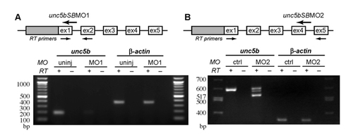
Knockdown of unc5b decreases levels of wild-type mRNA. RT-PCR analysis of control and morphant embryos shows that 1 ng unc5bSBMO1 or 4 ng unc5bSBMO2 significantly reduce levels of wild-type unc5b mRNA compared with uninjected embryos or to 8 ng control MO. Diagrams (top) show partial unc5b genomic structure (gray box, 5′ UTR; white boxes, coding exons; not to scale), with positions of MO sequences and RT-PCR primers indicated. Agarose gel images (below) show RT-PCR results for unc5b message and β-actin as a loading control. RT+/-, with or without reverse transcriptase. unc5bSBMO1 appears to abolish wild-type mRNA, while unc5bSBMO2 reduces wild-type mRNA and yields two additional bands. Sequencing of PCR products confirms that the upper band represents the wild-type product. The lower band corresponds to an in-frame deletion of amino acid residues 162-182, presumably reflecting a cryptic splice donor in exon 4. The middle band is a heteroduplex of the wild type and the alternate splice products. This deletion is in an Ig domain, which has been shown to be required for netrin binding (Kruger et al., 2004). DNA ladder sizes indicated in base pairs. |