- Title
-
Development of the proepicardial organ in the zebrafish
- Authors
- Serluca, F.C.
- Source
- Full text @ Dev. Biol.
|
The epicardium in the larval zebrafish heart expresses wt1. (A) Hematoxylin and eosin-stained section of a 4 dpf zebrafish embryo detailing the structure of the embryonic heart. The atrium (at) has a thinner wall than the ventricle (v) and valves are present between the two chambers (arrows). Myocardial nuclei are round while epicardial nuclei (arrowheads in the inset) appear elongated. (B) Whole-mount in situ hybridization of a 4 dpf embryo with the wt1 probe. The pronephric expression is marked with an asterisk (*) and cardiac staining (arrow) is clearly visible. (C) Frontal section of a 4 dpf wt1-stained embryo confirming that the wt1 expression is localized to the outer cardiac layer (arrow). Anterior is to the left in all panels. e: eye, y: yolk. EXPRESSION / LABELING:
|
|
Proepicardial organ lies in apposition to the myocardium of the embryonic heart. (A) A lateral view of the zebrafish heart at about 50 hpf with clusters of spherical cells seen using differential interference contrast optics nestled between the atrium and the yolk (arrow) and adjacent to the sinus venosus (arrowhead). Anterior is to the right. (B) Ventral view of the heart (50 hpf) with a cluster of cells attached to the outer ventricular wall (arrow). Anterior is at the top of the panel. at: atrium, v: ventricle. Movies of these embryos can be found in the supplemental information section. |
|
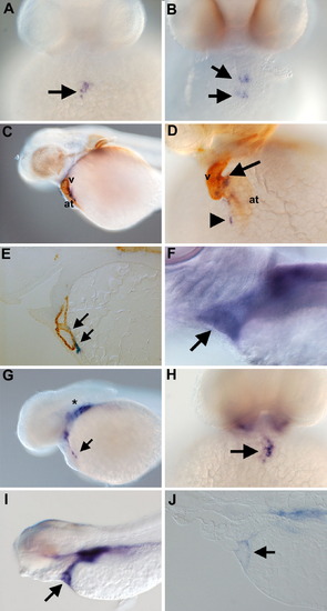
Expression of wt1 and tcf21 in the proepicardial organ. (A) Face view of a 40 hpf embryos stained with wt1. The arrow indicates the wt1-positive proepicardial organ. (B) At 48 hpf wt1 staining is visible as clusters (arrows). (C and D) Lateral and face views of whole-mount embryos co-stained with wt1 (blue) and the anti-myosin heavy chain antibody MF20 (brown). The wt1 clusters localize to the either the atrioventricular junction (arrow in panel D) or the sinus venosus (arrowhead in panel D). (E) Section of 72 hpf embryo stained with wt1 (blue) to label the developing epicardium and MF20 (brown) to label the heart muscle. A streak of wt1-positive cells is visible along the dorsal aspect of the heart (arrows). (F) At 120 hpf, the wt1-positive epicardium surrounds the heart (arrow). (G) Lateral and (H) face views of a 40 hpf embryo stained for tcf21 expression. tcf21 transcripts are located in a cluster of cells between the myoc>-positive epicardium surrounds the heart (arrow). (G) Lateral and (H) face views of a 40 hpf embryo stained for tcf21 expression. tcf21 transcripts are located in a cluster of cells between the myocardium and yolk (arrows) in an identical pattern to wt1. Strong expression of tcf21 is also visible in the developing arches (asterisk in panel G). (I) At 96 hpf, the heart is completely surrounded by tcf21-positive cells (arrow). (J) Sagittal section of a tcf21-stained embryo. As with wt1, the tcf21-positive layer at 96 hpf is confined to the outer epicardial layer (arrow). at: atrium, v: ventricle. |
|
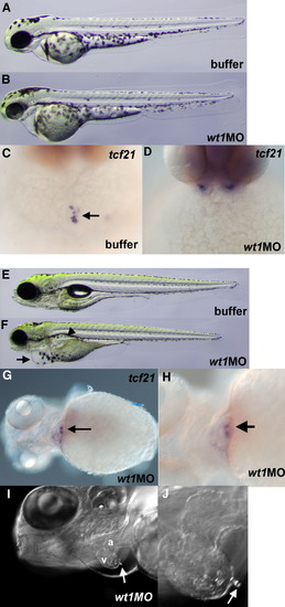
wt1 is required for development of the PEO. Live 48 hpf embryos injected with buffer (A) as a control or with wt1MO (B). A slight pericardial edema is observed in the wt1MO embryos. tcf21 staining at 48 hpf of (C) buffer-injected or (D) wt1MO-injected embryos. The PEO is clearly visible in the control embryos (arrow) but is completely lacking in wt1MO embryos (127/127, 100%). (E) Control and (F) wt1MO-injected embryos shown at 4 dpf. wt1MO embryos display pericardial edema (arrow in panel F) as well as edema in the pronephros (arrowhead). (G) At 4 dpf, a fraction of the embryos (22/68, 32%) have tcf21-positive cells adjacent to the heart (arrow). (H) Same embryo as panel G but shown at higher magnification with the wt1-positive cells (arrow) nestled between the heart and yolk as observed in 48 hpf wild-type embryos. (I) Live view of a 4 dpf wt1MO embryo using DIC optics. The ventricle (v) and atrium (a) are marked and an arrow points to a PEO-like cluster on the surface of the ventricular myocardium. (J) Higher magnification view of the embryo in panel I. |
|
Bifid hearts are each associated with their own proepicardial organ. Face views of buffer-injected (A) and milMO-injected (B) embryos stained with MF20 against myosin heavy chain (brown) and wt1 to mark the PEO (blue). A PEO cluster is visible at near the AV junction in buffer-injected embryos (arrow in panel A) while each heart in the milMO embryo is associated with a cluster of wt1-positive PEO (arrows in panel B). (C) Dorsal and (D) dorsolateral views on embryos injected with casMO. The nephric wt1 expression is indicated by the arrowheads and lies ventral to the anterior somites stained with MF20 (brown). Large, diffuse clusters of wt1 cells can be seen in the lateral positions (arrows). Dorsal views are shown because these cells fail to reach their more ventral positions next to the heart, seen here below the plane of focus in brown (asterisk). Anterior is to the left in panels C and D. |
|
pandora/spt6 is required for the PEO lineage. (A) Buffer-injected control embryos shown as a live 48 hpf embryo, (B) stained with wt1 (blue)/MF20 (brown) to mark the PEO and myocardium respectively, and with tcf21 (C) as a second marker of the PEO. (D) spt6MO embryos shown as live 48 hpf embryos. Note the extensive pericardial edema (asterisk) and tail extension defects (arrow). The PEO is not visible either by (E) wt1 or (F) tcf21 staining. Myocardium is still present, however, as evidenced by the MF20 positive tissue in the wt1/MF20 double-stained spt6MO embryos (arrowhead in panel E). (G) hegMO embryos display an enlarged heart (arrow) but relatively a relatively normal PEO as assayed by (H) wt1, with two wt1-positive clusters at the characteristic positions (arrowheads) and (I) tcf21 expression. (J) sihMO embryos shown live at 48 hpf. Staining with (K) wt1 and (L) >wt1-positive clusters at the characteristic positions (arrowheads) and (I) tcf21 expression. (J) sihMO embryos shown live at 48 hpf. Staining with (K) wt1 and (L) tcf21 reveals no effect of heart function on the formation of the PEO (arrowhead in panel K). Anterior is to the left in all panels except B, C, I, K and L which are face views with dorsal at the top of the panel. |
|
The cell polarity genes has and nok are required for proper PEO formation. (A) hasMO embryos shown live at 48 hpf. (B) hasMO embryo stained with MF20 (brown) and wt1 (blue) displays a midline heart but bilateral clusters of wt1 cells (arrows). (C) An example of a hasMO embryo displaying scattered wt1-positive cells (arrows) adjacent to the heart. (D) 48 hpf embryos injected with nokMO displaying characteristic pericardial edema. (E and F) nokMO embryo at 48 hpf stained with wt1 (blue) and MF20 (brown). Note the midline heart in but the bilateral spots of wt1 expression (arrows in panel E) and scattered wt1-positive cells in panel F. |
|
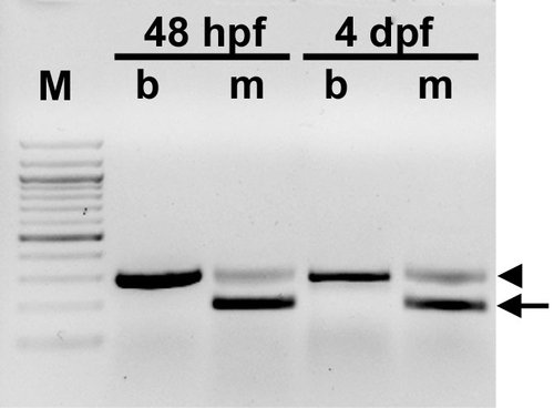
RT-PCR analysis of wt1MO embryos. RNA from buffer (b) injected and (m) wt1MO injected embryos was used as a template in a RT-PCR designed to amplify a portion of the wt1 messenger RNA at 48 hpf or 4 dpf. Most of the mRNA amplified in the morpholino injected embryos is aberrantly spliced yielding a smaller PCR product (arrow) than in wild type (arrowhead). M: 100 base pair ladder. Some wild type message is still present in the morpholino-injected embryos. PHENOTYPE:
|
|
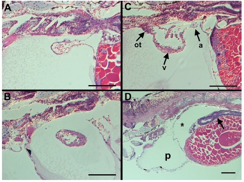
Histological analysis of 4 dpf wt1MO embryos. The hearts of embryos injected with the wt1 Morpholino are morphologically normal. (A) Atrium (arrow) from a wt1MO embryo. The chamber is single-layered and continuous with the sinus venosus. (B) Ventricle from a wt1MO embryo. Note the thicker ventricular wall as compared to the single-layered atrium in (A). (C) Sagittal section through a wt1 morpholino-injected embryo showing both the single-layered atrium (a) and thicker ventricular layer (v) and the outflow tract (ot) in a single section. (D) Lower magnification view of a wt1MO embryo with the pericardial edema marked by (p) and coelomic edema marked by and asterisk. The gut appears to be well differentiated (arrow). All sections shown are in the sagittal plane with anterior to the left, scale bar is 100 microns in all panels. |
Reprinted from Developmental Biology, 315(1), Serluca, F.C., Development of the proepicardial organ in the zebrafish, 18-27, Copyright (2008) with permission from Elsevier. Full text @ Dev. Biol.


