
- Title
-
Visualization of monoaminergic neurons and neurotoxicity of MPTP in live transgenic zebrafish
- Authors
- Wen, L., Wei, W., Gu, W., Huang, P., Ren, X., Zhang, Z., Zhu, Z., Lin, S., and Zhang, B.
- Source
- Full text @ Dev. Biol.
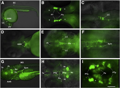
GFP expression pattern of ETvmat2:GFP embryos. Photos were taken under a fluorescence microscope in lateral (A, C, G), dorsal (B, E, F, H) and ventral views (D), or under a confocal microscope in dorsal view (I), rostral to the left. (A) A 24 hpf embryo showing the first GFP-positive (GFP+) neurons that appear in the nervous system; (B) a 30 hpf embryo showing GFP+ neurons in posterior tuberculum (PT) and their early axons projecting both anteriorly and posteriorly (asterisks), and some faintly labeled neurons in the paraventricular organ (Pa); (C) a 36 hpf embryo showing GFP+ neurons in the locus coeruleus (LC) located in the ventral lateral region of rhombomere 1; (D) a 54 hpf embryo showing the GFP+ arch-associated neurons (AAN) rostral to the heart; (E) a 60 hpf embryo with GFP+ neurons in the LC, the raphe nuclei (Ra) and the pretectum (Pt); (F) a 72 hpf embryo with GFP+ sympathetic neurons (Sym) under the artery; (G, H) 5 dpf embryos showing GFP+ neural groups in the central and peripheral nervous systems; (I) a 4 dpf embryo showing GFP+ neurons in PT and Pa, with the PT neurons sending out prominent single lateral neurites (asterisks). Scale bar: 25 μm. AAN: arch-associated neurons; Hc: caudal hypothalamus neural cluster; Hi: intermediate hypothalamus neural cluster; LC: locus coeruleus; MO: medulla oblongata neural cluster; Pa: neural cluster of paraventricular organ; Pt: pretectal neural cluster; PTa: anterior group of the posterior tubercular neurons; PTp: posterior group of the posterior tubercular neurons; Ra: raphe nuclei; SC: spinal cord; Sym: sympathetic neurons; Te: telencephalic neurons. Arrowheads in panels A and H: GFP-positive neurons in midbrain. |
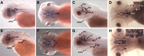
Comparison of vmat2 and th expression patterns. (A–H) Whole-mount in situ hybridization detecting vmat2 (A–D) and th (E–H) expression in 48 hpf (A, B, E, F) and 4 dpf (C, D, G, H) embryos. (A, C, E, G) Lateral views, (B, D, F, H) dorsal views, rostral to the left. Vmat2-reactive neurons in ventral diencephalon can be divided into anterior group of posterior tuberculum (PTa), posterior group of posterior tuberculum (PTp) and group of paraventricular organ (asterisk), and there is a gap (white arrow) between PTa and PTp; (H) catecholaminergic clusters in ventral diencephalon of 4 dpf embryos can be divided into several groups from anterior to posterior: population 1 (between ventral thalamus and posterior tuberculum), population 2 (anterior group of posterior tuberculum), population 3 (paraventricular organ), population 4 (posterior group of posterior tuberculum) and populations 5 and 6 (between posterior tuberculum and hypothalamus); there is also a gap (white arrow) between populations 2 and 4. AAN: arch-associated neurons; DC: diencephalic catecholaminergic cluster; DV: diencephalic vmat2-expression cluster; HyV: hypothalamic vmat2-expression cluster; LC: locus coeruleus; MC: medulla catecholaminergic cluster; PTa: anterior group of the posterior tubercular neurons; PTp: posterior group of the posterior tubercular neurons; PtC: pretectal catecholaminergic cluster; PtV: pretectal vmat2-expression cluster; Ra: raphe nuclei; Sym: sympathetic neurons. |
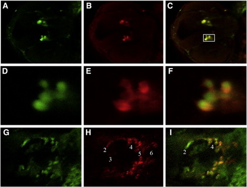
Double immunofluorescence staining for detection of GFP and endogenous Th. Simultaneous immunofluorescence staining was generated using a polyclonal anti-GFP antibody (A, D, G) and a monoclonal anti-Th antibody (B, E, H) for 30 hpf (A–F) and 3 dpf (G–I) ETvmat2:GFP embryos. Composite photos were made from stacked confocal images, focusing on catecholaminergic neurons of the ventral diencephalon. Merged pictures of Th and GFP staining are shown in panels C and F. Higher magnification images of the posterior tubercular neurons (D–F) correspond to the framed region in panel C. EXPRESSION / LABELING:
|
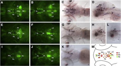
Effect of MPTP on zebrafish embryos. ETvmat2:GFP embryos untreated (A–D) or treated with 20 μg/ml (E–H) or 40 μg/ml (I–L) MPTP from 24 hpf to 5 dpf were examined at the last day. Photos were taken under a fluorescence microscope in ventral views (A, B, E, F, I, J), and whole-mount embryos after th in situ hybridization are in dorsal (C, G, K) and lateral (D, H, L) views, with rostral to the left. The neural clusters of ventral diencephalon and hypothalamus at a deep focal plane reduced significantly after MPTP exposure (E, I), while neural clusters located more dorsally and appeared at a shallow focal plane were almost unaffected (F, J). Similar reduction of ventral diencephalic and pretectal catecholaminergic clusters after MPTP treatment was observed with TH staining (G, H, K, L). Note that TH population 1 and 6 (G) and the pretectal cluster (G, H) were already eliminated after exposure to 20 μg/ml MPTP. (M) A schematic diagram representing the MPTP-sensitivity of different monoaminergic neuronal groups. 1, 2, 4, 5, 6: ventral diencephalic TH populations; DC: diencephalic catecholaminergic cluster; Hc: caudal hypothalamus neural cluster; Hi: intermediate hypothalamus neural cluster; HyV: hypothalamic vmat2-expression cluster; LC: locus coeruleus; Pa: neural cluster of paraventricular organ; Pt: pretectal neural cluster; PtC: pretectal catecholaminergic cluster; PtV: pretectal vmat2-expression cluster; PTa: anterior group of the posterior tubercular neurons; PTp: posterior group of the posterior tubercular neurons; Ra: raphe nuclei; Te: telencephalic neurons. Hi, highly MPTP-sensitive; Mo, moderately MPTP-sensitive; Re, MPTP-resistant. EXPRESSION / LABELING:
|
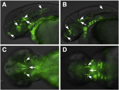
Effect of cyclopamine on ETvmat2:GFP embryos. Lateral (A, B) or dorsal (C, D) views of 48 hpf ETvmat2:GFP embryos after treatment with cyclopamine at 0 μg/ml (A, C) or 20 µg/ml (B, D) from 4 hpf to 48 hpf. Note that the raphe nuclei cluster (arrow) was abolished. Arrow: raphe nuclei cluster; arrowhead 1: locus coeruleus cluster; arrowhead 2: posterior tubercular cluster; arrowhead 3: telencephalic cluster; arrowhead 4: medulla oblongata cluster. EXPRESSION / LABELING:
|
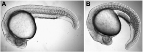
Morphologic effects of cyclopamine on zebrafish embryos. Lateral view of 24 hpf embryos after cyclopamine treatment at 0 μg/ml (A) or 20 μg/ml (B). |
Reprinted from Developmental Biology, 314(1), Wen, L., Wei, W., Gu, W., Huang, P., Ren, X., Zhang, Z., Zhu, Z., Lin, S., and Zhang, B., Visualization of monoaminergic neurons and neurotoxicity of MPTP in live transgenic zebrafish, 84-92, Copyright (2008) with permission from Elsevier. Full text @ Dev. Biol.