- Title
-
Zebrafish dlx2a contributes to hindbrain neural crest survival, is necessary for differentiation of sensory ganglia and functions with dlx1a in maturation of the arch cartilage elements
- Authors
- Sperber, S.M., Saxena, V., Hatch, G., and Ekker, M.
- Source
- Full text @ Dev. Biol.
|
(A) Expression of dlx2a in migrating neural crest cells at 13 somites (15 hpf) and 15 somites, neural crest streams are labeled with Roman numerals (inset; 16 hpf, dorsal view). Expression of (B) dlx2a and (C) dlx1a at 35 hpf. d, diencephalon; e, eye; h, hyoid arch; m, mandibular arch; ov, otic vesicle, t, telencephalon. Gill arches are numbered. Scale bar: (A) 100 μm, (B, C) 50 μm. (C–E) Histograms depicting the percentage of embryos and larvae showing abnormal head development following morpholinos injection. Embryos and larvae were evaluated based on proper head growth and/or normal chondrogenesis visualized by alcian blue staining (see Fig. 2). (C) Embryos injected with dlx2aMO and (E) embryos injected with dlx1aMO + dlx2aMO were assessed at 24 hpf. (D) Embryos injected with dlx1aMO were assessed at 120 hpf. Embryos failing to undergo proper extension and convergence due to the injection or general poor development due to possible toxi3E were assessed at 24 hpf. (D) Embryos injected with dlx1aMO were assessed at 120 hpf. Embryos failing to undcity were removed prior to assessment. Numbers of animals assessed for each morpholino and concentration of the injected morpholino (mM) are indicated within or below each bar, respectively. EXPRESSION / LABELING:
|
|
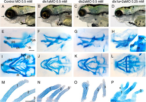
dlx1a and dlx2a are required for proper head outgrowth and arch cartilage patterning. (A–D) Lateral views of head morphology in 5-day-old larvae that received the (A) control MO, 0.5 mM; (B) dlx1aMO, 0.5 mM; (C) dlx2aMO, 0.25 mM; (D) an equal combination of dlx1aMO + dlx2aMO 0.25 mM each. (E–P) Cartilage elements stained with alcian blue. (E–H) are lateral views; (I–L) are ventral view. The inset in panel H is a lateral oblique view showing loss of the interhyal by fusion of hyosymplectic and ceratohyal elements (arrowhead). (M–P) Dissected ceratobranchial arch elements, numbered. cb, ceratobranchials; ch, ceratohyal; eth, ethmoid plate; hys, hyosymplectic; ih, interhyal; M, Meckel′s cartilage; pq, palatoquadrate. Arrows (A–D) indicate position of the mouth; asterisk in panel P indicates site of lateral branching. (A–P) Anterior is to the left. Scale bar in panel A: 250 µm; panels E and I: 100 μm; panel H inset, and M: 50 μm. PHENOTYPE:
|
|
dlx2a is necessary for survival of a subset of neural crest cells migrating to the pharyngeal arches. Crestin expression at 13 hpf in (A) Control (un-injected) embryo compared with, (B) an embryo injected with dlx2aMO, 0.25 mM. (B, inset) crestin expression at 15 hpf in dlx2aMO-injected embryo (0.25 mM). (C, D) Acridine orange staining of (C) control un-injected and (D) dlx2aMO-injected (0.5 mM) embryos at 14 hpf. (E, F) TUNEL-labeled transverse sections of a control (un-injected) 16 hpf embryo and an embryo injected with 0.25 mM dlx2aMO, (G, H) TUNEL labeling (black) and crestin (red) whole-mount in situ hybridization at 19 hpf. (G) Control (un-injected) embryo compared with (H) dlx2aMO injected embryo, 0.25 mM. Arrowheads in panels A and B indicate reductions of rostral crestin expression. Arrowheads in panels C, D, H indicate sites of cell death. (G, H) crestin labeled neural crest streams are labeled with Roman numerals. Anterior is to the left. cns, central nervous system; e, eye; m, mesoderm; nc, neural crest; ov otic vesicle; y, yolk. Scale bar: (A, G) 100 μm, (E) 50 μm. EXPRESSION / LABELING:
PHENOTYPE:
|
|
Zebrafish dlx2a is necessary for survival of a subset of cranial neural crest cells at later stages of pharyngeal arch development. TUNEL labeling was performed on 32 hpf embryos that received (A, A′) the control MO, 0.5 mM; (B, B′) dlx1aMO, 0.5 mM; (C, C′) dlx2aMO, 0.25 mM; (D, D′) dlx1aMO + dlx2aMO, 0.25 mM each. Anterior is to the left. e, eye; ov, otic vesicle. Boxed area (A) delineates region of interest (ROI) magnified in the associated A′–D′ panels. Arrows indicate cells labeled by TUNEL. Scale bar (A–D): 100 μm; (A′–D′): 50 μm. (E) Histogram depicting the average number of cells undergoing apoptosis within the ROI. Numbers of cells undergoing apoptosis, within the boxed area, were derived by counting and averaging cells from 10 embryos for each morpholino injected. PHENOTYPE:
|
|
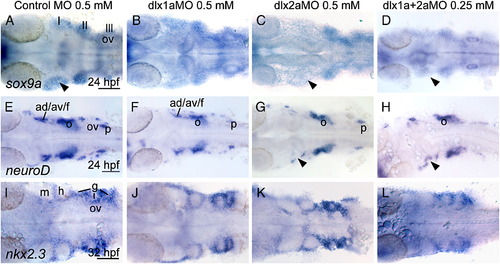
Effects of dlx1a, and dlx2a loss of function on arch markers. (A, E, and I) Control MO, 0.5 mM; (B, F and J) dlx1aMO, 0.5 mM; (C, G and K) dlx2aMO, 0.25 mM; (D, H, and L) dlx1aMO + dlx2aMO, 0.25 mM each. Expression of (A–D) sox9a, (E–H) neuroD and (I–L) nkx2.3. All panels show dorsal view of flat mounted embryos with anterior to the left. Arrowheads indicate sites of reduced or lost expression. Roman numerals indicate cranial neural crest streams. g, gill arches; ad/av/f, anterodorsal/anteroventral lateral line/facial placode/ganglia; h, hyoid; m, mandibular arch; o, octavel/statoacoustic ganglia precursors; ov, otic vesicle; p, posterior lateral line placode/ganglion. Scale bar: 100 μm. |
|
Loss of dlx2a but not of dlx1a function perturbs barx1 and goosecoid (gsc) expression in the developing second arch (a2) at 48 hpf. (A, E) Control MO 0.5 mM; (B, F) dlx1aMO 0.5 mM; (C, D) dlx2aMO 0.5 mM; (D, H) dlx1aMO + dlx2aMO 0.25 mM each. (A–D) barx1 expression; arrow indicates the intermediate region. (E–H) gsc expression in lateral views, arrowhead indicates location of the dorsal expression domain in the morphants. a1, first arch; a2, second arch; d, dorsal expression domain; v, ventral expression domain. Scale bar: 50 μm. EXPRESSION / LABELING:
|
|
Comparison of zn-5 staining in control and dlx1a, and dlx2a, and dlx1a/dlx2a morphants. Whole-mount immunohistochemical staining of 34 hpf embryos with the zn-5 antibody. Control embryos (A) are compared with embryos that were injected with (B) dlx1aMO, 0.5 mM; (C) dlx2aMO, 0.5 mM; (D) dlx1aMO + dlx2aMO 0.25 mM each. All panels are lateral views with anterior to the left. gV, trigeminal ganglia; gX, vagus ganglia; ov, otic vesicle; gill arches are underlined. Scale bar: 50 μm. EXPRESSION / LABELING:
|
|
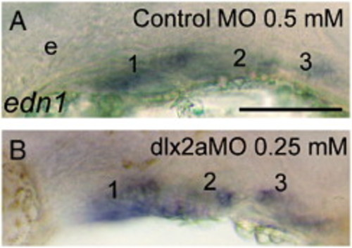
Expression of the edn1 in 24 hpf embryos that were injected with (B) 0.25 mM dlx2aMO embryos is similar to that of embryos that received the control MO (0.5 mM). Anterior is to the left. e, eye. Scale bar: 100 μm. EXPRESSION / LABELING:
|
|
Expression of hand2 in the pharyngeal arches of 34 hpf embryos that were injected with: (A) 0.5 mM control MO; (B) 0.5 mM dlx1aMO; (C) 0.5 mM dlx2aMO; (D) dlx1aMO + dlx2aMO, 0.25 mM each. Reductions are observed in embryos that received dlx2aMO or dlx1aMO + dlx2aMO. Arrowheads indicate hand2 expression Lateral views with anterior to the left. m, mandibular arch; h, hyoid arch; gill arches are numbered. Scale bar: 50 μm. EXPRESSION / LABELING:
|
Reprinted from Developmental Biology, 314(1), Sperber, S.M., Saxena, V., Hatch, G., and Ekker, M., Zebrafish dlx2a contributes to hindbrain neural crest survival, is necessary for differentiation of sensory ganglia and functions with dlx1a in maturation of the arch cartilage elements, 59-70, Copyright (2008) with permission from Elsevier. Full text @ Dev. Biol.


