
- Title
-
Snail1a and Snail1b cooperate in the anterior migration of the axial mesendoderm in the zebrafish embryo
- Authors
- Blanco, M.J., Barrallo-Gimeno, A., Acloque, H., Reyes, A.E., Tada, M., Allende, M.L., Mayor, R., and Nieto, M.A.
- Source
- Full text @ Development

snail1b knockdown induced morphological defects at gastrulation and segmentation stages. (A) Two different morpholino antisense oligonucleotides were designed against adjacent sequences in the 5' region of the snail1b cDNA. (B,D) Embryos at mid-gastrulation and (C,E) at the 18-somite stage. Morpholino injection (3 ng/embryo) produced defects in extension movements at early embryonic stages that led to the shortening of the body axis at 30 hpf (compare H with I). (F,G,J) This phenotype was rescued by co-injection of snail1b mRNA. (K,L,N,O,P,R) snail1a but not snail1b was prominently expressed in the involuting and posterior mesoderm. (M,Q) Lateral views highlight the expression of snail1a in the posterior half of the embryo and snail1b in the anterior. The dotted lines indicate the position of the margin and the adaxial cells (P) and that of the prechordal plate (R). ac, adaxial cells; ame, anterior axial mesendoderm; cm, condensing mesenchyme; pm, paraxial mesoderm; tbm, tail bud mesenchyme. PHENOTYPE:
|

snail1b knockdown does not affect mesodermal fate. (A-D) The axial and paraxial mesoderm form normally in snail1b morphants at tail bud stages (tb), as observed by the expression of ntl and paraxial protocadherin (papc), respectively. (E,F) MyoD expression at the 18-somite stage shows that knockdown of snail1b does not prevent the formation of somites. EXPRESSION / LABELING:
PHENOTYPE:
|
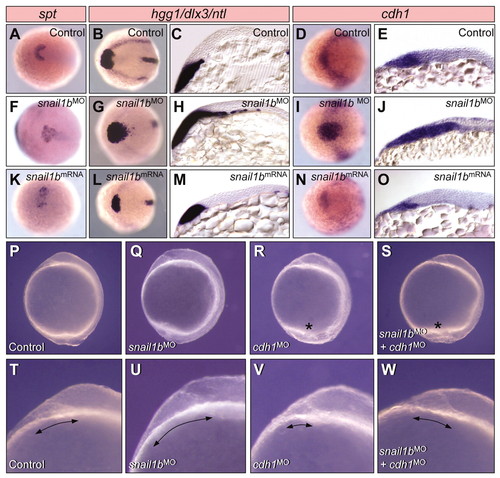
Snail1b modulates extension movements during gastrulation. (A,F,K) Knockdown of snail1b caused the posterior expansion of the spt expression domain, whereas overexpression of snail1b mRNA produced anterior compression. Similar effects were observed on the expression of hgg1 (B,G,L). (D,I,N) The expression of cdh1 follows that of hgg1 in the prechordal plate of control, as well as in snail1b morphants or overexpressing embryos. (C,H,M) Parasagittal sections obtained from hgg1-hybridised embryos reveal the position of the presumptive prechordal plate cells. Sections obtained from cdh1-hybridised embryos (E,J,O) confirm the similarity between hgg1 and cdh1 expression. (P-S) Increased cellular adhesion seems to account for the defects in extension movements in snail1b morphants, since the phenotype can be ameliorated by impairing cadherin activity with the injection of cdh1 MOs. The morphology of the polster is altered in the snail1b morphants (Q), although this effect can be reverted by the injection of cdh1 MOs (S). Asterisks indicate the defective posterior region where cells detach from the embryo both in the cdh1 (R) and in the double morphants (S). (T-W) Higher magnification of the images shown in P-S at the level of the prechordal plate to allow a better visualisation of the polster. The arrows indicate the territory it occupies along the anteroposterior axis. All embryos in this figure are at tail bud stages. |
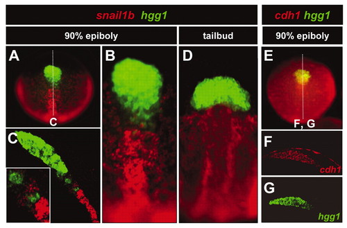
snail1b expression is excluded from the hgg1 expression domain within the axial mesendoderm. (A). Dual fluorescent in situ hybridisation demonstrates that snail1b expression is excluded from the hgg1-positive prechordal plate cells. The domains occupied by the two populations have ill-defined borders (B). (C) Sagittal sections of the embryo shown in A (dotted line) reveal the intermingling of the adjacent cell populations. Inset: border region at higher magnification. (D) At the tailbud stage, the cells have sorted out and a sharp boundary is defined. (E-G) The hgg1-positive prechordal plate cells also express cdh1 transcripts. EXPRESSION / LABELING:
|
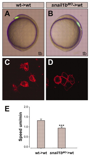
Migratory behaviour of snail1b morphant cells in wild-type hosts. (A,B) Lateral views of wild-type live embryos after transplantation at the shield stage of 10-20 axial mesendodermal cells from wild-type (A) or snail1b morphant (B) fluorescein dextran-labelled embryos. (C,D) Membrane-tagged RFP-labelled grafts from wild-type (C) or snail1b morphant (D) embryos on wild-type hosts at 70% epiboly stage allows the visualisation of differences in individual cell morphology: wild-type cells show protrusions whereas snail1b morphant cells form cohesive clusters and show an epithelial-like morphology. (E) Speed of wild-type and snail1b morphant axial mesendodermal cells in wild-type hosts (P<0.001). In each embryo, the distances of three individual cells were measured (wt→wt, n=7; mo→wt, n=5) PHENOTYPE:
|
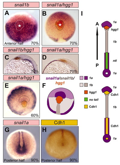
The axial mesendoderm is subdivided into different territories with complementary snail1 and E-cadherin (Cdh1) expression. (A-H) Expression of snail1 genes and that of E-cadherin protein. (A,B) At 70% epiboly, snail1a-expressing cells surround the anterior limit of the hgg1- expressing domain and snail1b transcripts are absent from the hgg1-expressing prechordal plate region. (C,D) These expression domains are better observed in sections. (E) The domain of strong snail1a expression surrounding the hgg1-expressing cells is already present at 60% epiboly. (F) Diagram depicting the domains of snail1a, 1b and hgg1 expression in the anterior half of the embryo. (G,H) snail1b and E-cadherin (Cdh1) expression in the posterior half of an embryo at 90% epiboly. Note the complementary domains of snail1b and cadherin expression (compare E with F). (I) Diagram showing the relationship between the expression of snail1 genes and those of hgg1 and ntl. Also note the inverse correlation between snail1 and Cdh1 expression. EXPRESSION / LABELING:
|
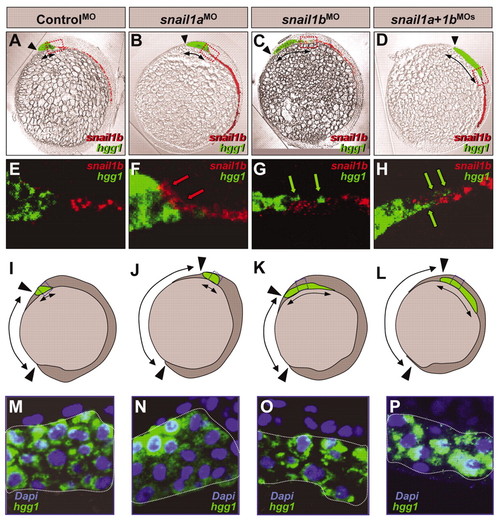
Snail1a and Snail1b cooperate in the directed anterior migration of the axial mesendoderm. (A-D) Morphological appearance of control embryos (A), and of embryos injected with snail1a MOs (B), with snail1b MOs (C) or with both (D). The expression of hgg1 (green) and snail1b (red) is superimposed. Note the abnormal position and/or shape of the prechordal plate. The arrows mark the territory occupied by the polster. (E-H) High-magnification confocal microscopy images of the same embryos at the regions indicated with a red square in A-D. Red arrows indicate the aberrant position of groups of snail1b-expressing cells, and green arrows that of hgg1-expressing cells. (I-L) Diagrams summarising the phenotypes observed in the snail1 morphants. (J) As previously described, the injection of snail1a MOs altered the posterior position of the prechordal plate (shown in green, with the extent indicated by a double-headed arrow). (K) snail1b morphants developed an elongated prechordal plate, which reached the normal anterior position. (L) Co-injection of snail1b and snail1a MOs produced a compound phenotype in the prechordal plate, which adopted an elongated shape and was situated at an aberrant posterior position. Black arrowheads indicate the anterior and posterior limits of the embryo. The double-headed arrows on the outside of the embryos mark the distance between the embryo limits. (M-P) hgg1 in situ hybridisation and DAPI (nuclei) staining show the cell density in the regions indicated with a dotted blue square in I-L. All embryos in this figure are at tail bud stages. |

The specificity of morpholino activity. The specificity of the morpholino activity was checked by GFP immunohistochemistry after co-injection of the morpholino and a plasmid vector encoding the complementary sequence of the morpholino in front of the EGFP sequence. When this construct and MO1 were co-injected, the translation of EGFP was impaired. EGFP production was tested by immunohistochemistry with a specific antibody (Molecular Probes) and visualized with the ABC Kit (Pierce). |

snail1b and no tail (ntl) expression domains. The posterior limit of snail1b expression in the axial mesendoderm coincides with the anterior limit of no tail (ntl) expression. EXPRESSION / LABELING:
|
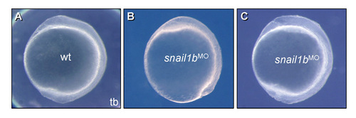
Wild-type and snail1b morphants at the end of gastrulation. (A) Wild type. (B) An embryo in which the hypoblast layer has reached the anterior position and formed the polster, but that has failed to complete epiboly. The prechordal plate was abnormally enlarged. (C) When embryos complete epiboly, the abnormal shape of the prechordal plate is more apparent. PHENOTYPE:
|