
- Title
-
Convergence and extension movements affect dynamic notochord-somite interactions essential for zebrafish slow muscle morphogenesis
- Authors
- Yin, C., and Solnica-Krezel, L.
- Source
- Full text @ Dev. Dyn.
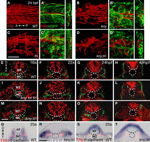
kny;tri double mutants are defective in slow muscle migration. A-D: Lateral views of the trunk region of the embryos stained with the slow muscle marker F59 antibody. Confocal z-stacks were collected and merged into one focal plane. A'-D': Left panels show the lateral views of the same embryos as shown in A-D, but only one single focal plane of the z-stacks is presented. The embryos are labeled with mGFP to highlight the morphology of the myotome and individual muscle fibers. Right panels show the confocal 3D reconstructed transverse sections. E-P: Localization of the slow muscle cells between the 16-somite stage (17 hpf) and 48 hpf as revealed by F59 antibody staining. SYTO59, which labels the nuclei, shows the embryonic structures. Q-T: m- and n-cadherin RNA expression (blue) and the positions of slow muscle cells revealed by F59 antibody labeling (pink) at the 25-somite stage (21.5 hpf). E-T: Transverse sections of the second or third somite. A, anterior; D, dorsal; NC, notochord; NT, neural tube; P, posterior; V, ventral. Scale bars = (A-D, A'-D', Q-T) 50 μm, (E-P) 20 μm. EXPRESSION / LABELING:
PHENOTYPE:
|
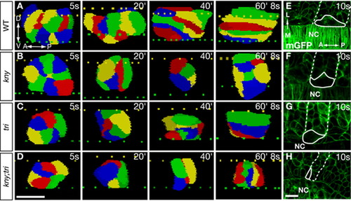
The kny;tri adaxial cells fail to undergo proper shape changes that precede the lateral migration. A-D: 3D reconstruction of the adaxial cells within the third somite (see Experimental Procedures section). Lateral views. Individual cells are colored for clarity. Dashed lines denote the dorsal (yellow) and ventral (green) surfaces of the notochord. E-H: Dorsal views of the embryos expressing mGFP, one adaxial cell (solid line) and the corresponding somitic boundaries (dashed line) are highlighted. A, anterior; D, dorsal; L, lateral; M, medial; NC, notochord; P, posterior; V, ventral. Scale bars = 20 μm (A-H). PHENOTYPE:
|
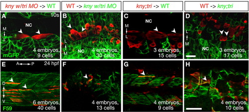
Kny and Tri act cell non-autonomously during slow muscle morphogenesis. A-H: Confocal images of the host embryos expressing mGFP (A-D) or stained with F59 antibody (E-H). Transplanted donor cells are labeled with Rhodamine dextran (red). The white arrowheads highlight several representative donor cells. A, B, E, F: Transplantation experiments using kny embryos injected with tri MO as donors (A, E) or hosts (B, F). C, D, G, H: Transplantation analyses using kny;tri double mutants as donors (C, G) or hosts (D, H). A-D: Dorsal views of the host embryos at the 10-somite stage (14 hpf). E-H: Lateral views of the host embryos at 24 hpf. A, anterior; D, dorsal; P, posterior; L, lateral; M, medial; NC, notochord; V, ventral. Scale bars = 20 μm (A-D), 50 μm (E-H). |
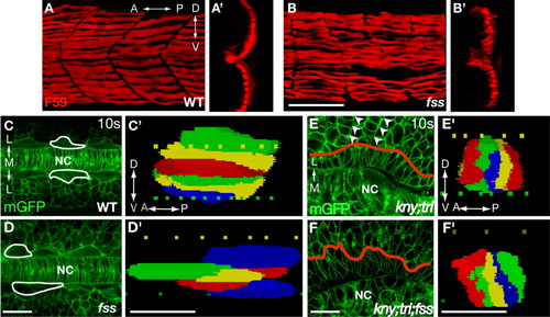
Somitic boundaries are dispensable for the shape changes and lateral migration of the adaxial cells. A, B: Lateral views of embryos stained with F59 antibody. A', B': 3D reconstructed transverse sections of the slow muscle fibers shown in A and B. C-F: Dorsal views of the embryos expressing mGFP. In C and D, white lines outline the selected adaxial cells. In E and F, red lines show the boundary between the adaxial cells and the lateral somitic cells. Arrowheads in E mark the somitic boundaries, which are missing in the kny;tri;fss embryos (F). C'-F': 3D reconstruction of the adaxial cells within the third somite at the 10-somite stage (14 hpf). Lateral views. A, anterior; D, dorsal; L, lateral; M, medial; NC, notochord; P, posterior; V, ventral. Scale bars = 50 μm (A,B, A',B'); 20 μm (C-F, C'-F'). PHENOTYPE:
|
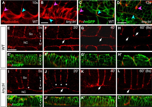
The adaxial cells in kny;tri double mutants exhibit prolonged contact with the notochord. A, B: F-actin distribution at the 10-somite stage (14 hpf) as revealed by phalloidin staining. C, D: Expression of Fn protein at the 12-somite stage (15 hpf). The embryos were co-labeled with mGFP to illustrate the morphology of the adaxial cells and the notochord. In A-D, the pink arrowheads point to the positions where the adaxial cells contact the anterior somitic boundary, whereas the blue arrowheads point to the opposite end of the adaxial cells. E-L: Time-course analyses of the Fn protein expression between the 5- and 8-somite stages. Arrows in F-H and J-K point to the expression of Fn protein at the notochord surface during the adaxial cell shape changes. E'-L': Confocal reconstructed sagital sections of the same embryos as shown in E-L, but were co-labeled with mGFP and Fn antibody. A-L: Dorsal views, anterior to the left. L, lateral; D, dorsal; M, medial; NC, notochord; So, somite; V, ventral. Scale bars = 20 μm (A-L, E'-L'). |
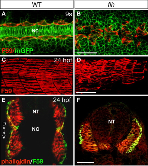
The presence of an intact notochord is not essential for slow muscle morphogenesis. A, B: Morphology of the adaxial cells in WT (A) and flh (B) mutant embryos at the 9-somite stage (13.5 hpf) as revealed by mGFP and F59 labeling. Notice the absence of the notochord and the aberrant positions of the adaxial cells in flh mutants (B). Dorsal views, anterior to the left. C,D: Confocal images of the slow muscle fibers stained with F59 antibody in WT (C) and flh mutants (D). Lateral views, anterior to the left. E,F: Transverse sections of the anterior myotomes stained with F59 antibody that labels the slow muscle cells, and phalloidin that recognizes both the fast and slow muscle fibers (Henry and Amacher,[2004]). A, anterior; D, dorsal; P, posterior; NT, neural tube; NC, notochord; V, ventral. Scale bars = 50 μm in A-F. PHENOTYPE:
|

Abnormal notochord properties impede the adaxial cell shape changes in kny;tri double mutants. A,B: Confocal images of the shield-depleted embryos at the 12-somite stage (15 hpf). The adaxial cells (arrows) are recognized based on F59 antibody staining. C,D: WT and kny;tri double mutant embryos expressing mGFP at the 12-somite stage. Insets show the notochord cells under higher magnification. E,F: Expression of Fn protein at the 12-somite stage (15 hpf). Dashed lines delineate the notochord-somite boundaries. Arrows in F point to the ectopic Fn expression inside the notochord in the double mutants. G,H: The presence of WT notochord cells suppressed the rotation defects of the double mutant adaxial cells lying next to them (white arrowheads). The WT donor cells are labeled with Rhodamine dextran (red). β-catenin antibody (green), which labels the cell membrane, and delineates the somite and notochord structures. A-H: Dorsal views, anterior to the left. NC, notochord; So, somite. Scale bars = 50 μm (A-F), 20 μm (G,H). PHENOTYPE:
|