- Title
-
Making sense of zebrafish neural development in the Minervois
- Authors
- Ghysen, A., Dambly-Chaudiere, C., and Raible, D.
- Source
- Full text @ Neural Dev.
|
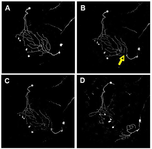
Imaging peripheral arbor re-innervation in vivo. Confocal projections showing dorsal views of two trigeminal axons, visualized with GFP, in a 54 hpf zebrafish embryo at several time points during a two-photon axotomy experiment. Anterior is to the bottom left. Images are each 420 microns across. (a) Twenty minutes before axotomy. (b) Approximately 20 minutes after axotomy. Yellow arrow points to site of axotomy. (c) Two hours after axotomy, the distal portion degenerates. (d) Robust regrowth is apparent 12 hours after axotomy. Figure provided by A Sagasti. |
|
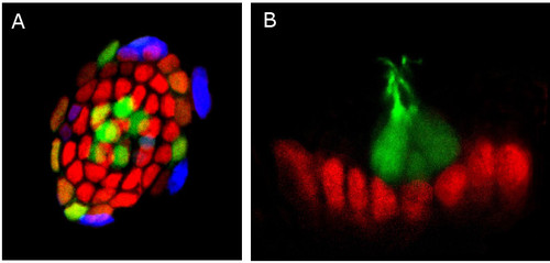
Distribution of Sox2 in lateral line neuromasts. (a) Confocal image of a zebrafish lateral line neuromast. Mantle cells and hair cells are labeled by GFP (green) in this transgenic line of zebrafish. Fluorescent immunostaining reveals expression of the neural progenitor marker protein Sox2 (red) and BrdU incorporation (blue). Cell division is occurring mostly in the periphery of the neuromast. (b) Confocal image of a zebrafish lateral line neuromast inmunostained to detect the neural progenitor marker protein Sox2 (red) and GFP (green) in hair cells. Note that these two markers do not overlap, suggesting that Sox2 is not present in differentiated cell types in neuromasts. Pictures provided by M Allende. |
|
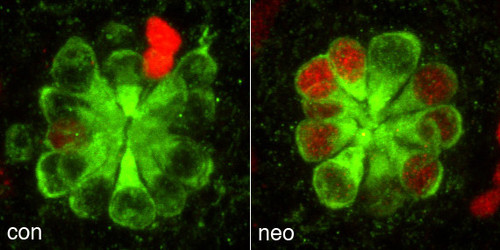
Control (con) and neomycin (neo) treated zebrafish lateral line neuromasts 72 hours after antibiotic treatment. Regenerated hair cells stained with anti-myosin VI antibody (green) are derived from proliferative precursors that incorporated BrdU (red). Figure provided by D Raible. |
|
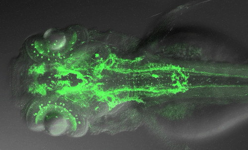
Projection on the horizontal plane of a Z-stack movie shown by J Schweitzer, illustrating a whole mount 3 dpf embryo labeled with an antibody against tyrosine hydroxylase. This picture was generated by S Ryu and W Driever. |


