
- Title
-
Requirements for Endothelin type-A receptors and Endothelin-1 signaling in the facial ectoderm for the patterning of skeletogenic neural crest cells in zebrafish
- Authors
- Nair, S., Li, W., Cornell, R., and Schilling, T.F.
- Source
- Full text @ Development
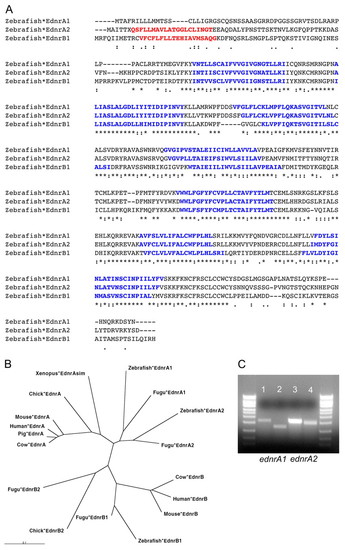
Characterization of zebrafish Ednras. (A) Alignment of zebrafish Ednras with Ednrb1. (B) The Ednr receptor family in vertebrates. (C) Splicing defects in Ednra morphants. PCR products of 1700 bp are seen in wild types (lane 1 ednra1, lane 3 ednra2), whereas morphants have one smaller product of 900 bp (lane 2, ednra1 MO) and 1500 bp (lane 4, ednra2 MO). |
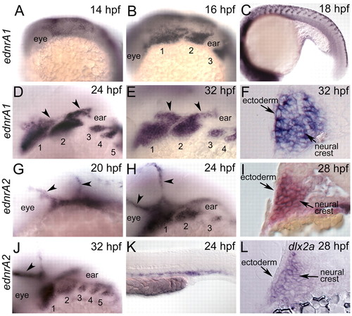
ednras are expressed in the pharyngeal arches. Whole-mount RNA in situ hybridizations in wild-type embryos; lateral views with anterior to the left (A-E,G,H,J,K). Transverse cryosections through the arches (F,I,L). (A) At 14 hpf, ednra1 is expressed in cranial NC. (B) At 16 hpf ednra1 marks three streams of cranial NC as well as NC in the trunk (18 hpf, C). (D) By 24 hpf, ednra1 is expressed throughout the entire DV extent of every arch. (E) Expression in NC cells is excluded from the endodermal pouches but is present in cranial ganglia (32 hpf, arrowheads in D,E). (F) Transverse cryosection at 32 hpf showing that ednra1 is expressed in both the NC and arch ectoderm. (G) ednra2 expression in cranial vasculature at 20 hpf (arrowheads in G,H,J). (H) ednra2 expression in NC cells of the arches at 24 hpf. (I) Co-labeling for dlx2a (red) and ednra2 (blue) showing both genes are expressed in NC cells throughout the arch and excluded from the ectoderm at 28 hpf. (J) At 32 hpf, ednra2 is expressed in NC, but like ednra1, is excluded from the pharyngeal endoderm. (K) ednra2 expression in trunk vasculature at 24 hpf. (L) dlx2a is expressed in NC cells throughout the arch but not the ectoderm at 28 hpf. EXPRESSION / LABELING:
|
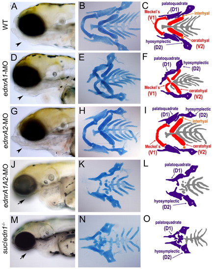
Ednra1 is required for jaw joints and patterns ventral cartilage redundantly with Ednra2. Lateral views of 96 hpf larvae (A,D,G,J,M); flat mounts of stained cartilage (B,E,H,K,N); schematics of cartilages (C,F,I,L,O). (A) The jaw protrudes beneath the eye in wild type (arrowhead). (D,G) In Ednra1 or Ednra2 morphants, the jaw is still prominent (arrowhead). (J) In Ednra1;Ednra2 double morphants, the jaw is absent (arrow), reminiscent of suc;edn1-/- (M, arrow). (B,C) Four distinct jaw elements are visible in the anterior two arches: Meckel's (red, V1), palatoquadrate (blue, D1), ceratohyal (red, V2) and hyosymplectic (blue, D2). (E,F) In Ednra1 morphants, all four develop but fuse. (H,I) Ednra2 morphants are indistinguishable from wild-type siblings, whereas in Ednra1;Ednra2 double morphants (K,L), and in suc;edn1-/- (N,O), ventral elements V1 and V2 are lost. PHENOTYPE:
|
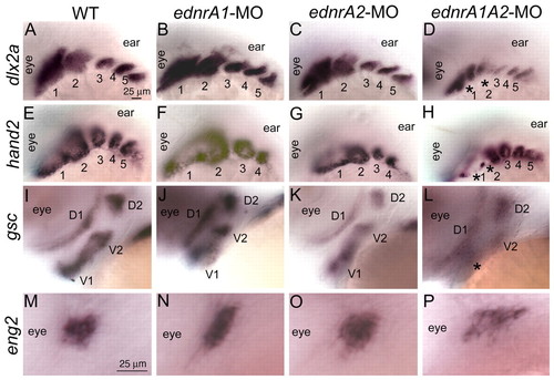
Ednras are required for patterning of ventral cranial NC. Whole-mount RNA in situ hybridization; 30 hpf dorsolateral views (A-H), lateral views (I-P). (A) dlx2a marks cranial NC cells along the DV axis of the arches, which remains unchanged in Ednra1 (B) or Ednra2 (C) morphants. (D) In Ednra1;Ednra2 double morphants, dlx2a is reduced in the ventral NC of arch 1 and 2 (asterisks). (E) hand2 marks rings of ventral NC in the arches, which remain in Ednra1 (F) or Ednra2 (G) morphants and are lost in Ednra1;Ednra2 double morphants (asterisks), except in small cell groups at arch borders (H). (I) At 44 hpf, gsc marks separate dorsal (D1, D2) and ventral (V1, V2) NC domains in the mandibular and hyoid arches, which remain unaffected in Ednra2 morphants (K). (J) In Ednra1 morphants, these domains remain distinct, although the distance between them is reduced. (L) In Ednra1;Ednra2 double morphants, gsc is dramatically reduced in the ventral mandibular (asterisk) and hyoid arches. (M) At 30 hpf, eng2 marks dorsal muscle precursors in the mandibular arch, which are unaffected in Ednra2 morphants (O). (N) In Ednra1 morphants eng2 extends ventrally. (P) In Ednra1;Ednra2 double morphants, eng2 expression spreads both along the DV and anteroposterior axes. Scale bars: 25 μm. |
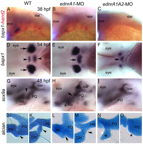
Ednra1 is required for joint formation in the arches. Whole-mount RNA in situ hybridization (A-I), dorsolateral views (A-C), ventral views (D-F), lateral views (G-I) and Alcian-Blue-stained, flat-mounted cartilage (J-O). (A) Two-color in situ hybridizations show bapx1 in joint precursors dorsal to hand2 in the ventral mandibular NC. (B) bapx1 expression is reduced in Ednra1 morphants. (C) In Ednra1;Ednra2 double morphants, both bapx1 and hand2 expression are abolished. (D) bapx1 bilaterally marks large jaw joint regions (arrow) as well as two smaller, ventral midline domains in the mandibular and hyoid arches (arrowheads). (E) In Ednra1 morphants, the bilateral domains of expression remain, presumably in cells surrounding the joints and a single, large domain persists at the ventral midline (arrow). (F) In Ednra1;Ednra2 double morphants, the two joint domains are severely reduced in addition to the ventral midline domain. (G) sox9a marks prechondrogenic NC cells in D1 (PQ) and V1 (Ms). A small gap in expression marks the joint between PQ and Ms (arrowhead). (H) In Ednra1 morphants, this gap is eliminated (arrow). (I) In Ednra1;Ednra2 double morphants, sox9a is expressed only in the remaining PQ condensation. (J) At 96 hpf, PQ and Ms are separated by a joint region (arrowhead) in the mandibular arch. (K) In the hyoid arch HS and CH are separated by the interhyal cartilage (arrowhead). (L,M) In Ednra1 morphants, these individual elements of both arches fuse (arrows). (N) In Ednra1;Ednra2 double morphants, a small remnant of Ms (asterisk) remains attached to PQ in the mandibular arch. (O) In the hyoid arch, CH is completely lost (asterisk). CH, ceratohyal; HS, hyosymplectic; Ms, Meckel's; PQ, palatoquadrate. |
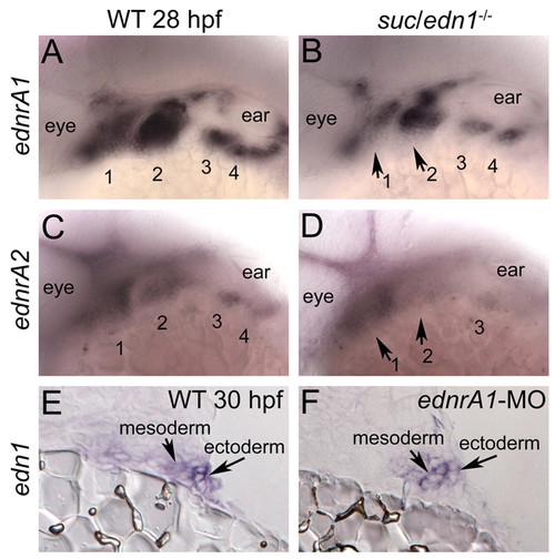
Interdependence of edn1 and ednrA in the ectoderm. Lateral views of whole-mount RNA in situ hybridizations (A-D). In B and D, mutants were identified as 25% of the embryos showing a distinct phenotype. (A) ednra1 is expressed in cranial NC cells of the arches. (B) In suc;edn1-/-, ednra1 is reduced in the ventral mandibular and hyoid arches (arrows). (C) ednrA2 is also expressed by cranial NC cells and is similarly reduced in suc;edn1-/- (D, arrows). (E) A transverse cryosection through the arch shows that edn1 is expressed in endoderm, mesoderm and ectoderm (arrows). (F) In Ednra1 morphants, edn1 is reduced in the pharyngeal ectoderm, although expression persists in the mesoderm (arrows). |
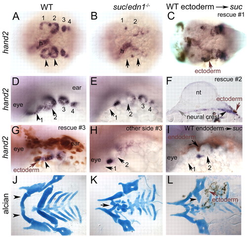
Facial ectoderm is a crucial functional source of Edn1 in the arches. Whole-mount RNA in situ hybridizations at 30 hpf (A,B,D,E), with immunohistochemistry for biotin-dextran (brown cells in C,F-I). Dorsal views (A-C), dorsolateral views (D,E,G-I), 96 hpf flat-mounted Alcian-Blue-stained cartilages (J,K) combined with immunohistochemistry for biotin-dextran (L). (A,D) hand2 expression in ventral cranial NC cells in wild type (arrowheads), which is lost in suc;edn1-/- (B,E, arrows), except in few cells at arch borders. (C) Rescue of hand2 in suc;edn1-/- by unilateral grafting of ectoderm on the left side (arrow). (F) Transverse cryosection through the arch shows unilateral rescue of hand2 (arrowhead) in a suc;edn1-/- embryo adjacent to grafted ectoderm (brown cells, arrow). The control side did not receive any donor ectoderm and shows no rescue of hand2 (gray arrow). (G) Another example of wild-type ectoderm (brown cells, arrow) rescuing hand2 (arrowheads). (H) Control side of embryo in G. (I) Wild-type endoderm (brown cells, arrow) did not rescue hand2. (J) Wild-type cartilages include the mandibular, hyoid and branchial elements including Meckel's cartilage (arrowhead). (K) Meckel's cartilage is reduced in suc;edn1-/- (arrow). (L) suc;edn1-/- mutant that received wild-type ectoderm shows unilateral rescue of the ventral hyosymplectic cartilage (arrowhead) adjacent to the grafted ectoderm (arrow, brown cells). h, heart; nt, neural tube. EXPRESSION / LABELING:
|
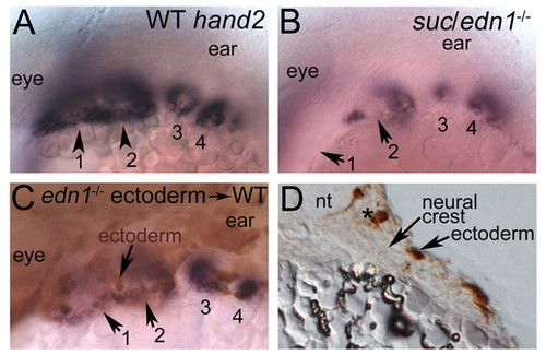
Edn1-deficient ectoderm recapitulates the suc/edn1-/-mutant phenotype. Whole-mount RNA in situ hybridizations (A,B) with immunohistochemistry for biotin-dextran (C,D). (A-C) Dorsolateral views. hand2 expression at 30 hpf (A) is lost in suc;edn1-/- mutants (B). (C) Wild-type embryos that receive ectoderm from suc;edn1-/- donors lose hand2 expression adjacent to the transplanted ectoderm (brown cells, arrow). (D) Transverse cryosection through the arches of the embryo in panel C show that donor cells are confined to ectoderm (arrow, brown cells). EXPRESSION / LABELING:
|