
- Title
-
Zebrafish atoh1 genes: classic proneural activity in the inner ear and regulation by Fgf and Notch
- Authors
- Millimaki, B.B., Sweet, E.M., Dhason, M.S., and Riley, B.B.
- Source
- Full text @ Development
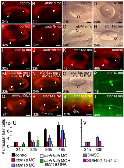
Requirement for atoh1 in hair cells in the ear and lateral line. All panels show dorsolateral views with anterior to the left and dorsal up. (A,B,E,F,I-N,Q-S) Pax2 antibody staining of otic hair cells (arrowheads) at the indicated times in control embryos (A,E,L), atoh1a morphant (I), atoh1b morphants (B,F), atoh1a;atoh1b double morphants (J,K), atoh1a;atoh1b double morphant co-injected with atoh1a mRNA (M,N) and embryos injected with atoh1a plasmid (Q-S). atoh1a plasmid stimulates production of supernumerary hair cells at 24 hpf (Q), but these are not maintained at 32 hpf (R), and instead displaced hair cells appear ventrally within subjacent mesenchyme, leaving gaps in the hair cell layer. An ectopic hair cell is revealed anterior to the otic vesicle by co-staining with Pax2a (red) and acetylated-tubulin (green) (S). (C,D,G,H,O) Otoliths produced in control (C), atoh1a morphant (G), atoh1b morphant (D) atoh1a;atoh1b double morphant (H) and atoh1a;atoh1b double morphant co-injected with atoh1a RNA (O). (P,T) Acetylated-tubulin staining of the lateral line and neuromasts (arrowheads) in atoh1b morphant (P) and atoh1a morphant (T) at 48 hpf. (U,V) The mean (± standard deviation) of Pax2-postive hair cells present in the utricle at the indicated times and under the indicated conditions. Sample sizes ranged from 15-35 embryos per time point. Scale bar: 15 μm. dhc, displaced hair cells; ehc, ectopic hair cell; o, otolith. |

Atoh1-dependent and -independent expression of atoh1 genes. Dorsolateral views (anterior to left) showing expression of atoh1a (A-C,G-I,M-O,S-U) and atoh1b (D-F,J-L,P-R,V-X) in control (A-F) atoh1a morphant (G-L), atoh1b morphant (M-R) and atoh1a;atoh1b double morphant (S-X) embryos at the indicated times. Expression of atoh1a at 32 hpf in mature hair cells and putative nascent hair cells is indicated in C. Arrowheads indicate observed or expected domains of otic expression. Inset in U shows a parasagittal section through the anterior atoh1a expression domain. Scale bar: 15 μm. hc, mature hair cells; n, nascent hair cells. EXPRESSION / LABELING:
|
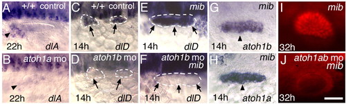
Interactions between atoh1 and the Delta-Notch pathway. (A,B) Expression of dlA at 22 hpf in a control embryo (A) and atoh1a morphant (B). (C-F) Expression of dlD at 14 hpf in a control embryo (C), atoh1b morphant (D), mib mutant (E) and mib mutant-atoh1b morphant (F). (G,H) mib mutants show expanded otic domains of atoh1b (G) and atoh1a (H) at 14 hpf. (I,J) Pax2 antibody staining at 32 hpf reveals supernumerary hair cells in a mib mutant (I) but no hair cells in a mib mutant co-injected with atoh1a MO and atoh1b MO (J). Arrowheads and arrows indicate otic regions. All images are dorsolateral views with anterior to the left. Scale bars: 30 μm in A,E,I-P; 15 μm in B-D,F-H. EXPRESSION / LABELING:
PHENOTYPE:
|

Heat-shock induction of dnSu(H). Expression of atoh1b at 13.5 or 14 hpf (A-C), atoh1b at 14.5 hpf (D-F) and Pax2 at 30 hpf (G-I), as seen in control embryos heat shocked at 10 hpf (A,D,G) or hsp70-dnSu(H) transgenic embryos heat shocked at 10 hpf (B,E,H) or 12 hpf (C,F,I). Images show lateral views with anterior to the left. Scale bar: 15 μm. EXPRESSION / LABELING:
PHENOTYPE:
|
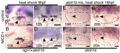
Heat-shock induction of NICD. (A-D) Expression of atoh1b and ngn1 at 11 hpf (A,B) and 12 hpf (C,D) in control embryos (A,C) or NICD-positive embryos (B,D) heat shocked at 9 hpf. Loss of ngn1 expression, which is non-overlapping with atoh1b, confirms effective NICD-induction. (E-H) Expression of atoh1a at 19 hpf (E,F) and 20 hpf (G,H) in atoh1b morphants without NICD (E,G) or with NICD (F,H) heat shocked at 18 hpf. Otic vesicles are outlined. Arrowheads mark otic expression domains. All are lateral views with anterior to the left. Scale bar: 15 μm. tg, trigeminal ganglion. EXPRESSION / LABELING:
PHENOTYPE:
|
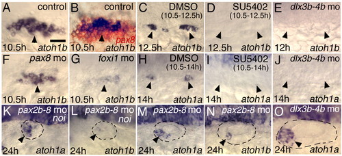
Inducers of early atoh1 expression. (A,B,F,G) Expression of atoh1b at 10.5 hpf in a control embryo (A,B), pax8 morphant (F) and foxi1 morphant (G). The specimen in B was double stained to reveal pax8 expression (red). (C,D) Expression of atoh1b at 12.5 hpf in embryos treated from 10.5-12.5 hpf with DMSO alone (C) or SU5402 in DMSO (D). (H,I) Expression of atoh1a at 14 hpf in embryos treated from 10.5-14 hpf with DMSO alone (H) or SU5402 in DMSO (I). (E,J,O) dlx3b;dlx4b morphants showing expression of atoh1b at 12 hpf (E) or atoh1a at 14 hpf (J) or 24h hpf (O). (K-N) Expression at 24 hpf of atoh1a (K,M) and atoh1b (L,N) in noi mutants injected with pax2b-pax8 MO (K,L), and in wild-type embryos injected with pax2b-pax8 MO (M,N). All are dorsolateral views with anterior to the left. Arrowheads indicate observed or expected domains of otic expression. Scale bar: 30 μm in A,B,F,G,K-O; 10 μm in C-E,H-J. EXPRESSION / LABELING:
|
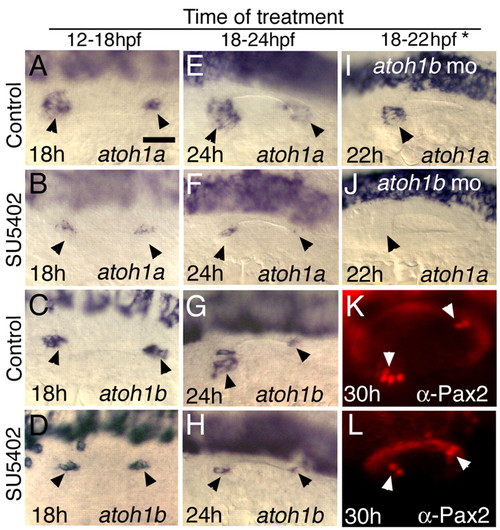
Stage-dependent requirements for Fgf. Embryos were treated with DMSO (control) or DMSO plus SU5402 for the indicated time intervals. (A-H) Expression of atoh1a in control and SU5402-treated embryos at 18 hpf (A,B) and 24 hpf (E,F), and expression of atoh1b in control and SU5402-treated embryos at 18 hpf (C,D) and 24 hpf (G,H). (I,J) Expression of atoh1a at 22 hpf in atoh1b morphants treated with DMSO (I) and DMSO and SU5402 (J). (K,L) Pax2 staining of hair cells at 30 hpf in embryos treated with DMSO (K) or DMSO and SU5402 (L). *, treatment for K, L from 18-24 hpf. All images are dorsolateral views with anterior to the left. Black arrowheads indicate otic expression. White arrowheads indicate sensory epithelia. Scale bar: 30 μm. EXPRESSION / LABELING:
|

Expression of macular genes. Expression of fgf3 (A,B) and fgf8 at 22 hpf (C,D), pax5 at 24 hpf (E,F) and pax2b at 30 hpf (G,H) in control embryos (A,C,E,G) and atoh1a;atoh1b double morphants (B,D,F,H). All panels show dorsolateral views with anterior to the left and dorsal up. Arrowheads indicate expression in sensory epithelia. Scale bar, 30 μm. EXPRESSION / LABELING:
|

Unillustrated author statements PHENOTYPE:
|