
- Title
-
Egr1 gene knockdown affects embryonic ocular development in zebrafish
- Authors
- Hu, C.Y., Yang, C.H., Chen, W.Y., Huang, C.J., Huang, H.Y., Chen, M.S., and Tsai, H.J.
- Source
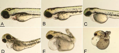
Morphological phenotypes of the wildtype and Egr1 morphants. Gross appearance of the wildtype (A) and Egr1 morphants (B: Grade 1, C: Grade 2, D: Grade 3, E: Grade 4, and F: Grade 5) at 72 h postfertilization. The grading criteria are defined in the Results section. The scale bar represents 100 μm in photo A, and is applicable to all photos. |
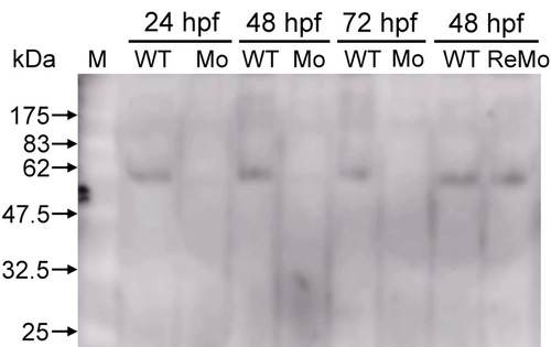
Western blot analysis of the Egr1 protein. A 55-kD Egr1 protein appears in wildtype (WT) embryos at 24, 48, and 72 h postfertilization (hpf), whereas the Grade-3 Egr1 morphants (Mo) do not exhibit this at the same periods. After Egr1 mRNA rescue, Egr1 protein appeared in the 48-hpf rescued morphants (ReMo). The leftmost lane represents the molecular weight marker (M). EXPRESSION / LABELING:
|
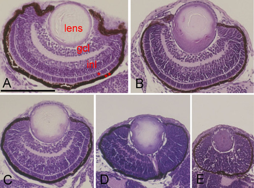
Histological examination of the wildtype and Egr1 morphants. Hematoxylin and eosin stained transverse sections of zebrafish eyes at 72 h postfertilization having A: wildtype, B: Grade 1, C: Grade 2, D: Grade 3, or E: Grade 4. Higher-grade morphants have smaller eyes and smaller lenses. Both the inner plexiform layer between the ganglion cell layer (gcl) and inner nuclear layer (inl) as well as the outer plexiform layer between the inl and outer nuclear layer (*) are thin. The outermost retinal pigment epithelium (**) layer is also thin and irregular. Scale bar represents 100 μm in photo A, and is applicable to all photos. |

Immunohistochemical staining of the wildtype and Egr1 morphants. Horizontal sections of zebrafish eyes at 72 h postfertilization with immunohistochemical stain for glutamate receptor 1 (A, B) and acetylated α-tubulin (C, D). Compared with the wildtype (A, C), retinal cells of the Grade-3 Egr1 morphant (B, D) arranged more compactly and disorderly. Significantly smaller areas of staining for both glutamate receptor 1 and acetylated α-tubulin appear in the morphant's retina. Scale bar represents 100 μm in panel A, and is applicable to all panels. |

Cryosection and immunostaining of the wildtype and Egr1 morphants. Cryosections of zebrafish eyes at 72 h postfertilization with zpr-1 immunostaining for photoreceptor cells. The wildtype (A) has markedly more labeled retinal cells in the outer nuclear layer (*) than the Grade-3 Egr1 morphant (B). The retinal pigmented epithelial layer (**) is also much thinner in the morphant. The scale bar represents 10 μm in photo A, and is applicable to photo B. |