
- Title
-
Zebrafish Trap230/Med12 is required as a coactivator for Sox9-dependent neural crest, cartilage and ear development
- Authors
- Rau, M.J., Fischer, S., and Neumann, C.J.
- Source
- Full text @ Dev. Biol.
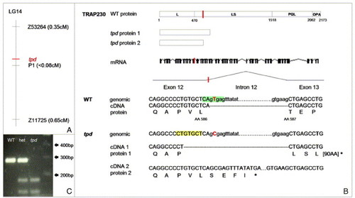
Molecular characterization of the tpd locus. (A) tpd maps to linkage group 14 (LG 14), between markers Z53264 and Z11725, close to marker P1. (B) The tpd mutation (marked in red) causes an alteration of the splice donor site of intron 12. Two defectively spliced transcripts are generated as a result of this mutation. cDNA1 is spliced at a cryptic splice site (shaded in yellow) within exon 12. This leads to a frame shift and a stop codon after 93 amino acids. cDNA2 contains unspliced intron 12, leading to a stop codon after 4 amino acids. (C) Restriction enzyme Est1 cuts genomic tpd but not WT DNA due to the point mutation in its recognition site. het: DNA from a heterozygous embryo. L: leucine-rich domain; LS leucine-and-serine-rich domain; PQL: proline-, glutamine-, and leucine-rich domain; OPA: glutamine-rich domain. |
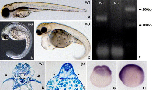
The overall Trap230 loss-of-function phenotype. (A, D, G, H) WT, (B, E) tpd, (C) Trap230 morpholino-injected embryos. (A–E) Embryos at 3 dpf. (D, E) Methylene blue-stained transverse sections at the level of the pectoral fin buds (arrows and asterisks). (F) Trap230 MO-injected embryos lack the RT-PCR product amplified from exons 26 and 27, since intron 26 fails to be spliced out. (G, H) trap230 mRNA expression. (G) Four-cell stage, animal pole to the top. (H) Three-somite stage, lateral view, dorsal to the top, anterior to the left. MO: Trap230 morpholino injected. EXPRESSION / LABELING:
|

Ear and pigmentation phenotypes in tpd mutants and Trap230 morphants. (A–G, J–L) Embryos at 4 dpf. (H, I) Embryos at 28 hpf. (A–E) Ear phenotypes; note partial reduction in tpd mutants (C), and complete absence in some Trap230 morphants (D). (F, G) Dark field microscopy, revealing iridophores (bright shiny cells), present in WT (F), but not in tpd mutants (G). (H, I) TUNEL staining, lateral view of embryos. (J–L) Trunk melanocytes (dark cells, arrows). (A, F, H, J) WT; (B, C, G, I, K) tpd mutants; (D, E, L) Trap230 MO-injected embryos. |
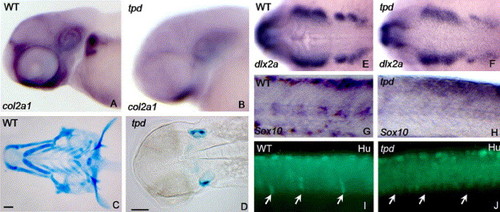
Neural crest and craniofacial phenotypes of tpd mutants. (A, C, E, G) WT; (B, D, F, H) tpd mutants. (A, B) Expression of col2a1 in the head at 68 hpf, lateral view. (C, D) Expression of dlx2a in the pharyngeal arches at 24 hpf, dorsal view. (E, F) Alcian blue-stained cartilages at 5 dpf, ventral view. (G, H) Sox10 Expression in the trunk region of wild type (G) and tpd mutants (H). Note the absence of ventrally migrating Sox10-expressing cells in tpd mutants. (I, J) Anti-Hu immunostaining at 3 dpf, lateral view of the trunk. Arrows indicate the position of DRGs. Note the absence of DRGs in tpd mutants. |

Sox9a and Sox9b expression in tpd mutants. (A, C, E, G) WT; (B, D) tpd mutants; (F, H) Trap230 MO-injected embryos. (A, B) Embryos at 10 hpf, animal pole to the top. (E, F) Embryos at 3-somite stage, dorsal view, anterior to the left. (C, D, G, H) Embryos at 24 hpf, lateral view, anterior to the left. (A–D) Expression of Sox9a. (E–H) Expression of Sox9b. Note that Sox9a and Sox9b expression is only weakly reduced in the absence of Trap230 (B, F). |

Trap230 is required as a coactivator for Sox9. (A, C, E, G, I, K, M, O) WT embryos. (B, D, F, H, J, L, N, P) tpd mutants. Embryos are at the 3-somite stage, dorsal view, anterior to the left. (A–D) foxd3 expression. (E–H, K, L) snai1b Expression. (I, J) Sox10 expression. (M–P) six3 expression. (C, G) WT embryo injected with Sox9b mRNA; (D, H) tpd mutant injected with Sox9b mRNA. (K) WT embryo injected with Sox9b-VP16 mRNA; (L) tpd mutant injected with Sox9b-VP16 mRNA. (O) WT embryo injected with prdm1 mRNA. (P) tpd mutant injected with prdm1 mRNA. EXPRESSION / LABELING:
|

Ear development in the absence of Trap230. (A, C, E, G) WT; (B, D, F, H) Trap230 MO-injected embryos. (A–D) Embryos at 3-somite stage, dorsal view, anterior to the left. (E, F) Tailbud stage embryos, dorsal view, anterior to the left. (G, H) Lateral views of the ear at 24 hpf. (A, B) Sox9b expression in the otic placodes; (C, D) Sox9b expression in the otic placodes. (E, F) pax8 expression in the otic placodes (arrows). (G, H) pax2.1 expression in the otic vesicle. EXPRESSION / LABELING:
|
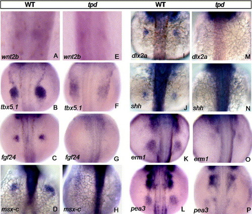
The pectoral fin phenotype of tpd mutants. All embryos shown are 24 hpf, dorsal views, anterior to the top. (A–D, I–L) WT; (E–H, M–P) tpd mutant embryos. (A, E) wnt2b expression. (B, F) tbx5.1 expression. (C, G) fgf24 Expression. (D, H) msx-c expression. (I, M) dlx2a expression. (J, N) shh expression. (K, O) erm1 expression. (L, P) pea3 expression. EXPRESSION / LABELING:
|

Unillustrated author statements |
Reprinted from Developmental Biology, 296(1), Rau, M.J., Fischer, S., and Neumann, C.J., Zebrafish Trap230/Med12 is required as a coactivator for Sox9-dependent neural crest, cartilage and ear development, 83-93, Copyright (2006) with permission from Elsevier. Full text @ Dev. Biol.