
- Title
-
BMP signaling restricts hemato-vascular development from lateral mesoderm during somitogenesis
- Authors
- Gupta, S., Zhu, H., Zon, L.I., and Evans, T.
- Source
- Full text @ Development

The I-SceI meganuclease system can be used to reduce mosaicism effectively in transient transgenic animals. In a series of experiments, it was found that the full and normal pattern of expression can be recapitulated using either the lmo2 or gata1 promoters in ~30% of the injected embryos. Shown are representative examples of embryos derived from fertilized eggs that had been injected with transgenes and the meganuclease. (A,B) Two independent examples using the I(lmo2;gfp)I transgene examined at the 10-somite stage. GFP expression is present throughout the two stripes of lateral mesoderm (arrows). Views are dorsal, anterior towards the left. (C) A similar embryo analyzed at 20 hpf, with GFP expression throughout the ICM (arrow) and also anterior lateral mesoderm (arrowhead). The view is lateral, anterior towards the left. (D) A representative embryo, viewed as in C, derived from eggs injected with the I(gata1:gfp)I construct at 20 hpf. GFP expression is restricted to the ICM (arrow). |

Using I-SceI-mediated transgenesis the expression of a dominant-negative BMP receptor can be targeted effectively to lateral mesoderm during somitogenesis. Shown are representative embryos processed for in situ hybridization using an antisense RNA probe specific for the Xenopus δ transgene (δbr). Embryos were either injected with the I(lmo2;δBR)I transgene (A-C) or represent uninjected sibling controls (D-F). Transgene expression is not detected in lateral mesoderm prior to somitogenesis, e.g. at the tailbud stage (A,D). However, at the three-somite stage (B,E) or the eight-somite stage (C,F), signal for the δbr transcript is detected throughout the two stripes of rostral and caudal lateral mesoderm (arrows in B and C), consistent with the normal Lmo2 expression pattern. Using this Xenopus-derived RNA probe all embryos (whether transgenic or not) showed a low level of non-specific background staining (indicated by the arrowhead in A). |

Initial specification of hemato-vascular progenitors is not affected by the lmo2:δBR transgene. Shown are representative sibling embryos that are either control (A) or transgenic for the lmo2:δBR transgene B). Embryos were fixed and processed by in situ hybridization to detect the expression of scl transcripts. In all cases, the initial pattern of scl is normal, as indicated here by the two stripes of lateral mesoderm (arrows) at the two-somite stage. EXPRESSION / LABELING:
|
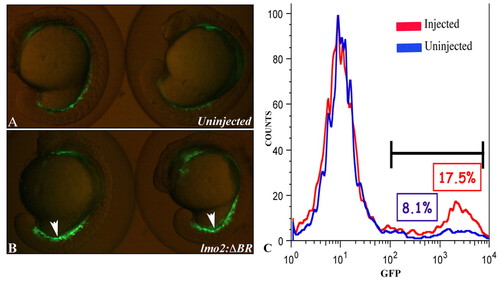
Expression of a dominant-negative BMP receptor in lateral mesoderm results in enhanced hematopoiesis. Two representative embryos are shown in A and B; views are lateral, anterior towards the top, with embryos still in their chorions. (A) Control embryos from the gata1:gfp transgenic line demonstrate the normal expression pattern of GFP in the ICM. (B) Embryos from the gata1:gfp transgenic line that had been injected at the one-cell stage with the I(lmo2δBR)I transgene and meganuclease. GFP expression is substantially increased in the ICM, compared with control embryos (arrowheads). (C) Dissociated cells were collected at the 16- to 17-somite stage from batches of control and transient transgenic embryos and analyzed by FACS to score quantitatively the numbers of GFP+ cells. Shown are results from one representative experiment, although the data were comparable in three independent experiments. Compared with control embryos (blue) the I(lmo2δBR)I transgenic embryos (red) show more than double the normal amount of GFP+ hematopoietic cells. |

Expression of a dominant-negative BMP receptor in lateral mesoderm results in enhanced expression of early hemato-vascular markers. (A-C) Representative embryos analyzed by in situ hybridization for expression of gata1. Views are flat-mounted dorsal, anterior towards the top. (A) Control uninjected embryo (B) Embryo that had been injected with I(lmo2:δBR)I. (C) Embryo that had been injected with I(lmo2:cre)I. gata1 expression is markedly increased in lateral mesoderm both laterally (arrowheads) and in the caudal region of the embryos expressing δBR, compared with the controls (arrow). (D-G) Representative embryos analyzed for myeloid markers. Views are ventral, anterior towards the top (D,E) or lateral, anterior towards the left (F,G). Compared with uninjected control embryos (D,F), the embryos transgenic for I(lmo2:δBR)I (E,G) have increased expression levels of transcripts for l-plastin (E) and mpo (G) (arrowheads). (H) Control and (I) a corresponding representative I(lmo2:&deltaBR)I transgenic embryo analyzed for expression of the endothelial marker flk1. The embryos expressing the mutant receptor show enhanced levels of flk1 in the posterior tail (arrowheads). Views are lateral to the tail region, anterior towards the left. (J,K) Representative embryos from the fli:gfp transgenic line used to evaluate vascular development. Compared with control embryos (J), embryos expressing the mutant receptor in lateral mesoderm display dilation of the vasculature in the ICM region (arrowheads). Views are lateral, anterior towards the left. EXPRESSION / LABELING:
|

Both Lmo2+ and Gata1+ hematopoietic cells are restricted by BMP signaling. Representative embryos are shown that are either control (A,C,E,G) or derived from fertilized eggs injected with either I(lmo2:δBR)I (B,D) or I(gata1:δBR)I (F,H), and analyzed for gene expression by in situ hybridization. The markers tested are as indicted: (A,B) scl; (C,D) lmo2; (E,F) gata1; (G,H) flk1. Expression of both scl and lmo2 is enhanced by expression of the mutant receptor in Lmo2+ cells (arrowheads in B and D), while gata1 (the hematopoietic marker; arrowheads) but not flk1 (the endothelial marker; arrows) is enhanced when the receptor is expressed in Gata1+ cells (E-H). Views are flat-mounted dorsal, anterior towards the left (A-F) or lateral, anterior towards the left (G,H). EXPRESSION / LABELING:
|
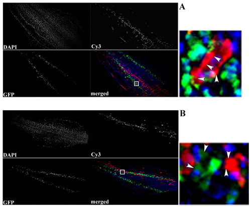
Excess Bmp4 in lateral mesoderm restricts the number of neighboring hematopoietic cells. Donor cells were derived from embryos that had been injected either with rhodamine-conjugated dextran alone (A) or also with RNA encoding Bmp4 (B). Cells were transplanted into developing embryos that are transgenic for the gata1:gfp reporter. A and B show representative chimeric embryos that contain similar numbers of transplanted (red) cells localized to lateral mesoderm. Embryos were also stained with Hoechst (blue) to distinguish each cell (whether red, green or unlabeled). For each red cell in lateral mesoderm, the number of GFP+ hematopoietic cells in the immediate neighborhood was determined. When donor cells express excess Bmp4, there is a decreased ratio of green (host hematopoietic) to red (donor) cells (data compiled in Table 2). A and B illustrate a single representative embryo for each case, showing the three channels (DAPI, Cy3 and GFP) and a merge, with a small insert indicated in the merged panel on the right. In these magnified panels, the presence of four red cells are indicated by arrowheads. In B, they are associated with a single green (Gata1+) cell, whereas multiple green cells are present in A. Views are flat-mounted dorsal, anterior towards the left. |

Hyperactive BMP signaling in lateral mesoderm restricts hemato-vascular development. Shown are representative sibling embryos that were either control (A,C) or transgenic for the lmo2:caBR transgene (B,D). Embryos were fixed at the 12-somite stage and processed by in situ hybridization to detect the expression patterns of the gata1 gene (A,B; marking committed hematopoietic cells) or the scl gene (C,D; marking hemato-vascular progenitors). The patterns (indicated in the caudal lateral mesoderm by arrows) are markedly repressed by expression of the hyper-activated receptor. EXPRESSION / LABELING:
|

BMP signaling enhances expression of the pronephric marker pax2.1 in lateral mesoderm. (A-E) Representative embryos derived from fertilized eggs that were control (A,D) or had been injected with the I(lmo2:δBR)I transgene to express the dominant negative receptor (B,E) or the I(lmo2:caBR)I to express the constitutively active receptor (C). Embryos shown in A-C were analyzed for expression of the pronephric marker pax2.1 (arrowheads) and co-stained for the expression of the midline mesoderm marker ntl as an internal control (asterisks). Embryos in D and E were processed sequentially to detect the expression of pax2.1 (purple) and gata1 (red). The magnified views in the side panels of D and E demonstrate that the gata1 pattern is expanded (white bars) at the expense of the pax2.1 pattern (black bars) in injected embryos. Views are flat-mounted dorsal, anterior towards the top. EXPRESSION / LABELING:
|