- Title
-
Control of cell migration in the zebrafish lateral line: Implication of the gene "Tumour-Associated Calcium Signal Transducer," tacstd
- Authors
- Villablanca, E.J., Renucci, A., Sapede, D., Lec, V., Soubiran, F., Sandoval, P.C., Dambly-Chaudiere, C., Ghysen, A., and Allende, M.L.
- Source
- Full text @ Dev. Dyn.
|
Localization of TACSTD protein. Zebrafish embryos were injected with myc fusion mRNAs, allowed to develop for 12 hr, and developed with an anti-myc antibody. A: Zebrafish tacstd-myc fusion mRNA was injected and embryonic cells were visualized under confocal microscopy; nuclei were labelled with propidium iodide (red) and the anti-myc antibody was detected with an Alexa fluorescent secondary antibody (green). B: Same as in A, but propidium iodide was not added. Note expression localized to membranes and to the perinuclear region. C: Injected mRNA corresponds to myc-tagged SKIP protein, a transcription factor localized to the nucleus. |
|
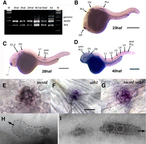
Developmental expression of tacstd. A: RT-PCR detection of tacstd message at 0, 6, 24, 48 haf, 6 daf, and adult (Ad) stages. Amplification of Wnt5a message is used as a positive control. The adult mRNA sample was contaminated with genomic DNA as amplification of genomic sequence produces a larger product (labelled as "genomic"). B-D: Whole mount in situ hybridization using a tacstd riboprobe. Embryos were processed for hybridization at 23 (B), 28 (C), and 48 haf (D). E-G: Neuromasts of in situ hybridized embryos. Probes used were tacstd (E), ath1 (F), or both probes (G). H,I: tacstd expression in the anterior lateral line (H) or posterior lateral line (I) primordia. Arrows indicate direction of migration. Dotted circles demarcate cells showing a rosette pattern, which is coincident with strong expression of tacstd. Expression to the left of the primordium in I is a recently deposited proneuromast. ALL, anterior lateral line; L1-5, posterior lateral line neuromasts; O1,2, otic neuromasts; OP, olfactory placode; OV, otic vesicle; PLL, posterior lateral line; prim, primordium. |
|
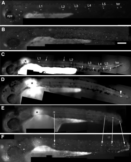
Embryonic posterior lateral line labeled by 4-Di-2-ASP, a marker of differentiated hair cells (A, B, and F) or by uncaging the primordium (C-E). A: Wild type embryo with the normal pattern of five neuromasts along the horizontal myoseptum (L1-L5) and two terminal neuromasts (ter). B: Morphant embryo where all PLL neuromasts are missing. C: Uncaging the primordium at the position marked by the asterisk in a wild type embryo reveals the neuromasts (L1-5 and ter) and interneuromastic cells (arrows). D: Uncaging in a morphant reveals that the primordium has reached the tip of the tail (arrowhead) but no neuromasts have been deposited. E,F: In a partial morphant where four clusters have been deposited, including a terminal one (E), each cluster has differentiated hair cells on the next day (F). PHENOTYPE:
|
|
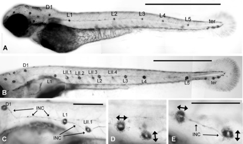
Alkaline phosphatase labeling of neuromasts. A: Normal pattern of neuromasts in a 2-day-old embryo. L1-L5 and the terminal neuromasts are derived form primI. D1, the first neuromast of the dorsal line, is derived from primII. B: Normal pattern in a 6-day-old larva. Consecutive primI neuromasts are connected by a thin trail of interneuromastic cells. Four additional neuromasts have been added by primII: LII.1-LII.4. The dorsal line comprises three additional neuromasts along the dorsal midline (out of focus). C: The trail of primI interneuromastic cells (INC) is continuous with the primI neuromast L1 but appears pushed away by the derivatives of primII (D1 and LII.1). D,E: The phosphatase activity reveals neuromast anisotropy, with the primI neuromasts being polarized along the antero-posterior axis while the primII neuromasts are polarized in a dorso-ventral direction (double arrows). Scale bars = 1 mm (A,B), 250 μm (C-E). |
|
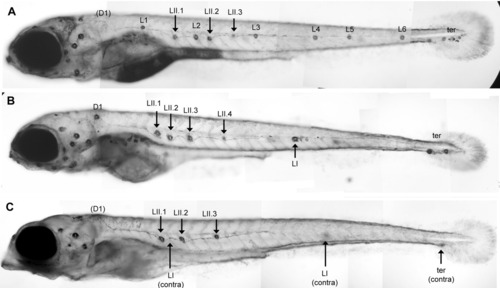
Effect of tacstd inactivation on secondary lateral line formation. A: A normal 6-day-old larva showing three primII neuromasts (LII.1, LII.2, and LII.3). B,C: Two morphant embryos showing a strong phenotype. B: One lateral and two terminal primI neuromasts are present. The primII neuromasts have formed normally (LII.1-LII.4). C: No primI neuromast is present on the focused side of the embryo; two lateral and one terminal primI neuromasts are present on the other side as indicated. This embryo also shows the stereotyped head defect present in about 30% of the embryos. Note that in A and C, the D1 neuromast has been dislodged during manipulation. This rarely happens and can easily be detected because a rim of labelled cells remains attached to the epidermis, as can be faintly seen at this low magnification in A. PHENOTYPE:
|

ZFIN is incorporating published figure images and captions as part of an ongoing project. Figures from some publications have not yet been curated, or are not available for display because of copyright restrictions. |


