
- Title
-
her1 and her13.2 are jointly required for somitic border specification along the entire axis of the fish embryo
- Authors
- Sieger, D., Ackermann, B., Winkler, C., Tautz, D., and Gajewski, M.
- Source
- Full text @ Dev. Biol.
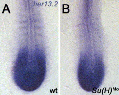
her13.2 expression in Su(H) morphants. (A) wt expression of her13.2 and (B) in Su(H) morphants (0.6 mM; 2 experiments, n = 65, 96.92% affected). Note the reduction of stripes might be rather a secondary effect due to loss of somite borders in this morphant. Embryos are between the 8 and 10 somite stage, dorsal view, flat mounted embryos, anterior to the top. EXPRESSION / LABELING:
|
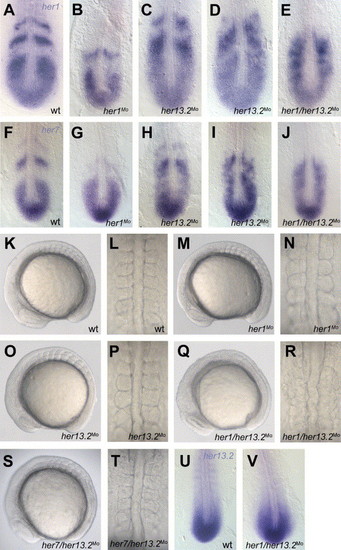
Effects of her1/her13.2 injections on somite borders and expression patterns. (A), (F) wild-type expression of her1 and her7, respectively. (B), (G) her1 and her7 expression, respectively, after her1 knockdown (0.6 mM; 2 experiments, n = 76 for each probe, 92.11% affected for her1, 94.74% affected for her7). her13.2Mo injection (0.6 mM; 2 experiments; n = 53 for her1, n = 43 for her7) leads in 73.58% of the embryos to a slightly altered her1 expression (C) and in 81.4% of the embryos to the same alteration in her7 expression (H). 9.43% of her1 stained embryos and 18.6% of her7 stained embryos show a full disruption of the her1 (D) and the her7 (I) expression, respectively. A full disruption of her1 expression (E) and her7 expression (J) is observed in her1/her13.2 double morphants (0.6 mM her1Mo + 0.6 mM her13.2Mo; 2 experiments, n = 85, 94.11% affected for her1, 95.29% affected for her7). (K), (L) somite morphology in wild-type embryos, (M), (N) in her1 morphants (0.6 mM; 2 experiments, n = 56, all embryos show almost wild-type morphology with slight border defects up to somite 3), (O), (P) in her13.2 morphants (0.6 mM; 2 experiments, n = 52, all embryos show wild-type morphology), (Q), (R) in her1/her13.2 double morphants (0.6 mM her1Mo + 0.6 mM her13.2Mo; 2 experiments, n = 92, 97.83% affected) and (S), (T) in her7/her13.2 morphants (0.6 mM her7Mo + 0.6 mM her13.2Mo; 2 experiments, n = 46, all embryos show wild-type morphology in the anterior somites). Note in the her1/her13.2 double morphants, the head appears slightly enlarged, with a dark lens primordium, the tailbud tip has a flattened appearance (Q), and the notochord is kinked (R) compared to the wild type (K). (U) Wild-type expression of her13.2 and (V) in her1/her13.2 morphants (0.6 mM her1Mo + 0.6 mM her13.2Mo; 2 experiments, n = 42, 92.86% affected (loss of stripes might rather be a secondary effect due to loss of somite borders)). (A–J), (U), and (V) dorsal view, flat mounted embryos, anterior to the top. (K), (M), (O), (Q), and (S) lateral view, anterior to the left. (L), (N), (P), (R), and (T) dorsal view of the anterior trunk somites, anterior to the top. EXPRESSION / LABELING:
|

Expression of myoD and somite border effects in the different morphants. (A) Wild-type expression of myoD, (B) in her1 morphants (0.6 mM; 2 experiments, n = 40, 95% show segmental myoD expression, which is slightly more diffuse in anterior somites), (C) in her13.2 morphants (0.6 mM; 2 experiments, n = 38, all embryos show segmental myoD expression) and (D) a full disruption of the myoD pattern in her1/her13.2 morphants (0.6 mM her1Mo + 0.6 mM her13.2Mo; 2 experiments, n = 57, 96.49% affected). Somite border disruption in (E) beatm98/deltaC mutants (van Eeden et al., 1996 and Jülich et al., 2005b), (F) her13.2Mo injections in beatm98/deltaC mutant background (0.6 mM; 2 experiments, n = 65, all embryos show bea phenotype), (G) Su(H) morphants (0.6 mM; 1 experiment, n = 40, 95% affected, showing the Su(H)Mo phenotype (Sieger et al., 2003)) and (H) Su(H)/her13.2 double morphants (0.6 mM Su(H)Mo + 0.6 mM her13.2Mo; 2 experiments, n = 52, 96.15% show Su(H)Mo phenotype). No enhanced effects could be observed in panels (F) and (H) when her13.2 morpholino was additionally injected compared to the single beatm98 mutant (E) or Su(H) morphant situation (G), respectively. (A–D) 8–10 somite stage embryos, flat mounted embryos, anterior to the top. (E–H) Whole-mount embryos, lateral view, anterior to the left. Stars in panels (E–H) mark the position of the somite borders. EXPRESSION / LABELING:
|

Disruption of early oscillations in her1/her13.2 morphants. Oscillations in gene expression of her1, her7, and deltaC are disrupted in her1/her13.2 morphants at bud stage. (A, E, and I) Wild-type expression of her1, her7, and deltaC, respectively. Dynamic gene expression can be found for her1 (B), her7 (F), and deltaC (J) in her1 morphants (0.6 mM; 2 experiments, n = 40 for each probe, all embryos show wild-type dynamics) since in a batch of embryos the whole variety of the pattern can be found (two patterns are shown each for wt, her1 morphant and her13.2 morphant situation, respectively). The same dynamic is seen in her13.2 morphants for her1 (C), her7 (G), and deltaC (K) (0.6 mM; 2 experiments, n = 45 for each probe, all embryos show wild-type dynamics). In her1/her13.2 morphants, a full disruption of cyclic her1 (D), her7 (H), and deltaC (L) expression is detected, and the whole batch of embryos shows only one pattern (0.6 mM her1Mo + 0.6 mM her13.2Mo; 2 experiments, n = 52 for each probe, 96.15% affected for her1, 94.23% affected for her7, and 92.3% affected for deltaC). (A–L) Bud stage embryos, whole mounts, dorsal view, posterior downwards. EXPRESSION / LABELING:
|
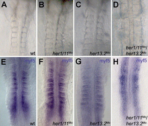
Effects of her1/11Mo and her13.2Mo injections on somite border formation in medaka. (A) Anterior somite borders in wild-type embryos, (B) in her1/11 morphants (0.2 mM, 2 experiments, n = 124, 97.58% embryos show wild-type morphology) and (C) in her13.2 morphants (0.2 mM, 5 experiments, n = 281, 98.9% embryos show wild-type morphology). (D) Anterior somite borders are completely absent in double injections (0.06 mM her1/11Mo and 0.2 mM her13.2Mo, 7 experiments, n = 267, 66.3% affected). Expression of the segmental marker myf5 (Elmasri et al., 2004) was monitored in (E) wild-type embryos, (F) her1/11 morphants (0.2 mM, 2 experiments, n = 82, 95% show segmental myf5 expression), (G) her13.2 morphants (0.2 mM, 3 experiments, n = 105, 97% show segmental myf5 expression) and (H) respective double morphants (0.06 mM her1/11Mo and 0.2 mM her13.2Mo, 3 experiments, n = 89, 96.6% show uniform myf5 expression). (A–G) 8–10 somite stage embryos, (A–D) living embryos, (E–G) flat mounted embryos, all in dorsal view, anterior to the top. |
Reprinted from Developmental Biology, 293(1), Sieger, D., Ackermann, B., Winkler, C., Tautz, D., and Gajewski, M., her1 and her13.2 are jointly required for somitic border specification along the entire axis of the fish embryo, 242-251, Copyright (2006) with permission from Elsevier. Full text @ Dev. Biol.