
- Title
-
Spatiotemporal expression of zebrafish keratin 18 during early embryogenesis and the establishment of a keratin 18:RFP transgenic line
- Authors
- Wang, Y.H., Chen, Y.H., Lin, Y.J., and Tsai, H.J.
- Source
- Full text @ Gene Expr. Patterns

Cytokeratin 18 (K18) expression during early embryonic stages. (A) At the one-celled stage, dorsal view. (B) At 8 h postfertilization (hpf), dorsal view. (C) Enlarged view of the same embryo in B. (D) At 10-hpf, dorsal view. (E) At 14-hpf, lateral view. (F) At 18-hpf, dorsal view. (G) At 20-hpf, lateral view. (H) Enlarged view of the same embryo in G; the arrow indicates the presumptive epithelial cells. (I) Detailed view of 48-hpf embryos; the arrow indicates the presumptive epithelial cells. |

Red fluorescent protein (RFP) expression of the transgenic fish lines during early embryogenesis. The F2 offspring were produced by mating F1 female from Tg(k18(2.9):RFP) line with wild-type males (A, B, D, E) or by mating the F1 males from the Tg(k18(2.9):RFP) with wild-type females (C). (A) At the one-cell stage. (B) At 14 h postfertilization (hpf). (C) At 14 hpf. (D) At 3 days postfertilization (dpf); the arrows and arrow head indicate two different shapes of epithelial cells. (E) At 4 dpf, the red signals were gradually abolished to the undetectable level (upper). However, some of them still displayed red fluorescence (bottom). (F) Observation under the light field. |
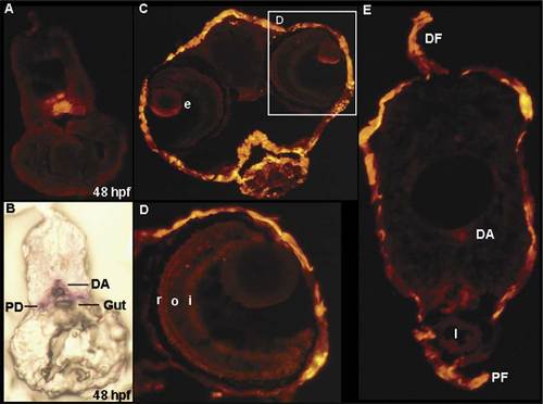
Cryosections of 48 h postfertilization (hpf) and 7 days postfertilization (dpf) larva from wild-type embryos and Tg(k18(2.9):RFP) transgenic line embryos. (A) Cryosection of the wild-type 48 hpf embryos, revealing that the endogenous k18 was expressed in the dorsal aorta, pronephric duct, and gut. (B) The comparable section from a 48 hpf k18(2.9):RFP transgenic embryo. (C) Head region. (D) Enlarged view of the same section in C. (D) Trunk section. DA, dorsal aorta; DF, dorsal fin; I, intestine; PD, pronephric duct; PF, pelvic fin; e, eye; i, inner plexiform layer; o, outer plexiform layer; r, rods and cones. |

(A–E) Red fluorescent protein (RFP) expression patterns of transgenic lines in the adult fish. The arrow indicates that no red fluorescence was observed when the scale was removed. AF, anal fin; CF, caudal fin; DF, dorsal fin; PF, pelvic fin. |
Reprinted from Gene expression patterns : GEP, 6(4), Wang, Y.H., Chen, Y.H., Lin, Y.J., and Tsai, H.J., Spatiotemporal expression of zebrafish keratin 18 during early embryogenesis and the establishment of a keratin 18:RFP transgenic line, 335-339, Copyright (2006) with permission from Elsevier. Full text @ Gene Expr. Patterns