- Title
-
Essential and overlapping roles for laminin alpha chains in notochord and blood vessel formation
- Authors
- Pollard, S.M., Parsons, M.J., Kamei, M., Kettleborough, R.N., Thomas, K.A., Pham, V.N., Bae, M.K., Scott, A., Weinstein, B.M., and Stemple, D.L.
- Source
- Full text @ Dev. Biol.
|
Morphological and molecular analysis of bal, gup and sly mutant embryos. Lateral views show wild-type (WT) (A, G), bal (D, K), gup (H) and sly (L) embryos at 3 day post fertilisation. Staining with a polyclonal antibody generated against mouse laminin 1 (α1β1γ1) detects expression pattern in 24 hpf WT (B) embryos. Laminin immunoreactivity in 24 hpf bal mutant embryos (E) is at wild-type levels within the posterior notochord (arrowhead), but severely reduced immunoreactivity is seen in the non-differentiated anterior notochord, and intersomitic boundaries. In 24 hpf gup (I) and sly (M) embryos, laminin 1 immunoreactivity is abolished. The developing vasculature is marked by expression of fli-1 mRNA in 24 hpf WT (C) and bal (F), gup (J) and sly (N) mutant embryos. EXPRESSION / LABELING:
PHENOTYPE:
|
|
Expression of lama1 mRNA and genetic mosaic experiments reveal that laminin α1 is supplied from non-notochordal tissues. Whole-mount in situ hybridisation for lama1 mRNA in wild-type zebrafish embryos at: 8-cell stage (A), shield stage (B), 5-somite stage (C, dorsal view), 15-somite stage (D), 25-somite stage (E) and 24 hpf (F, dorsal view of head; G, lateral view of trunk and tail). Embryonic shield transplantation from a Rhodamine-dextran labelled bal mutant donor (J), into wild-type host (H), gives rise to a secondary axis containing a rescued anterior notochord (red fluorescence) (I). Morphologically wild-type notochord in both the primary (host) axis and secondary (donor) axis are indicated with white arrowheads. |
|
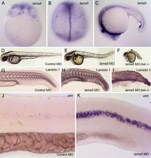
Expression and morpholino antisense knockdown of lama5 in zebrafish embryos. Whole-mount in situ hybridisation of lama5 mRNA in zebrafish embryos at 64-cell stage (A), tailbud stage (B, dorsal) and 20-somite stage (C, lateral). Injection of antisense MO directed against lama5 mRNA results in CNS disruptions and defective extension of the yolk sac by 28 hpf (E) compared to control MO injected (D). Notochord, however, appears fully differentiated, by morphology and laminin-1 immunoreactivity (H) compared to control MO-injected notochord (G). Embryos injected with lama5 MO display strong persistent expression of the early chordamesoderm marker, ehh (K), whose expression is extinguished by this stage in control MO-injected embryos (J). Injection of lama5 MO into bal mutant embryos results in severe disruption of many tissues (F), though laminin-1 immunoreactivity is retained (I). EXPRESSION / LABELING:
PHENOTYPE:
|
|
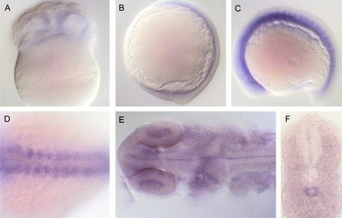
Expression of lama4 mRNA during zebrafish development. Whole mount in situ hybridisation shows localisation of lama4 mRNA in 16-cell (A), tailbud (B), 15-somite (C, lateral and D, dorsal view of trunk) and 24 hpf (E, dorsal view of head; F, section through trunk) wild-type embryos. |
|
Morpholino antisense knockdown of laminin α4 in bal mutants reveals redundant roles in notochord differentiation. Lateral views of 48 hpf control MO-injected embryos from a heterozygous bal cross showing a morphologically WT (A) and a bal mutant (B) (arrowhead; non-differentiated anterior notochord) embryo. From the same heterozygous bal cross, lama4 MO-injected embryos are also shown, ∼75% of embryos show a brain defect but no obvious notochord phenotype (C), whereas ∼25% of embryos have a severe notochord defect (D), with the entire anterior–posterior extent of the notochord disrupted. Whole mount laminin 1 antibody staining at 24 hpf shows expression of immunoreactivity in morphologically WT (E) and bal (F) mutant control MO-injected embryos. While ∼75% of lama4 MO-injected embryos have wild-type laminin 1 immunoreactivity (I), ∼25% (bal mutants) show severe loss of laminin 1 (J) (two independent experiments, total n = 136). Staining for expression of echidna hedgehog (ehh) at 24 hpf reveals the status of notochord differentiation in control MO-injected wild-type (G) and bal (H) mutant embryos. While lama4 MO-injected WT (K) embryos normally extinguish ehh expression, lama4 MO-injected bal mutant (L) embryos persistently express ehh. |
|
Laminin α1 and laminin α4 chains function redundantly during ISV sprouting. At 24 hpf, wild-type embryos (A) and bal mutant embryos injected with control MO (B) as well as wild-type embryos injected with lama4 MO (C) show normal fli-1 expression in both axial and intersegmental vessels. In contrast, bal mutants injected with lama4 MO (D) lack fli-1 staining of intersegmental vessels. |
|
Axial vessels are correctly specified but intersegmental vessels fail to properly sprout and extend in laminin α1 and α4 loss of function embryos. In situ hybridisation of embryos injected with control MO (A, C, E, G) or co-injected with lama1 MO and lama4 MO (B, D, F, H) stained for VE-cadherin (A, B), ephrinB2 (C, D), flt4 (E, F) or VEGF (G, H) expression. In double laminin α1 and α4 loss of function embryos, the dorsal aorta (red arrows) and posterior cardinal vein (blue arrows) properly express markers of endothelial differentiation (A, B) and arterial (C, D) and venous (E, F) identity. The proangiogenic factor VEGF is also comparably expressed in somitic tissue (black arrows) in double morphants (G, H). Multiphoton imaging of trunk vessels in Fli-EGFP transgenic embryos injected with control MO at 24 (I) or 48 hpf (J) or coinjected with lama 1 MO and lama4 MOs at 36(K) or 60 hpf (L). Primary intersegmental sprouts initiate but do not extend and show much less protrusive activity. All panels show lateral views of the mid-trunk region, with anterior to the left, except for panels G and H, which show dorsal views of the mid-trunk region in flat-mounted embryos, with the left side up. |
Reprinted from Developmental Biology, 289(1), Pollard, S.M., Parsons, M.J., Kamei, M., Kettleborough, R.N., Thomas, K.A., Pham, V.N., Bae, M.K., Scott, A., Weinstein, B.M., and Stemple, D.L., Essential and overlapping roles for laminin alpha chains in notochord and blood vessel formation, 64-76, Copyright (2006) with permission from Elsevier. Full text @ Dev. Biol.


