
- Title
-
Identification and comparative expression analysis of a second wt1 gene in zebrafish
- Authors
- Bollig, F., Mehringer, R., Perner, B., Hartung, C., Schafer, M., Schartl, M., Volff, J.N., Winkler, C., and Englert, C.
- Source
- Full text @ Dev. Dyn.
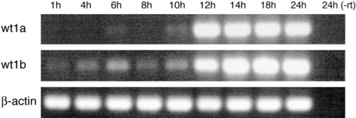
Temporal expression pattern of wt1a and wt1b during early zebrafish development. Analysis was performed by reverse transcriptase-polymerase chain reaction (PCR) using embryos of the stages indicated. Sizes of the amplified products were 520 bp for wt1a, 560 bp for wt1b, and 400 bp for β-actin, which was used as a reference. To verify the absence of genomic DNA contamination, an aliquot of the 24 hours postfertilization RNA sample was used for PCR without reverse transcription (-rt). h, hours postfertilization. |
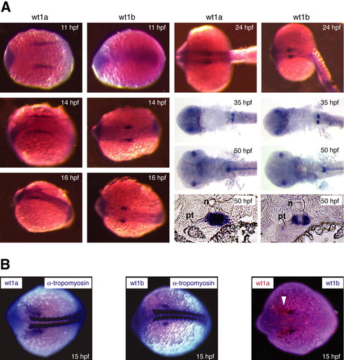
Expression of two wt1 genes in the developing zebrafish. A: Embryos were collected at the desired stages (hpf, hr postfertilization) and subjected to whole-mount in situ hybridization using wt1a and wt1b probes. Signals in the eyes at 35 and 50 hours postfertilization (hpf) as well as in the notochord at 50 hpf can also be observed using the respective sense controls (data not shown) and, therefore, are due to probe trapping. The two right bottom panels show adjacent cross-sections of the anterior glomerular region of a 50 hpf embryo that have been hybridized with wt1a and wt1b probes, respectively. n, notochord; pt, pronephric tubule. B: Double whole-mount in situ hybridizations were performed using probes against wt1a and the somite marker α-tropomyosin (left), wt1b and α-tropomyosin (middle), as well as against both wt1 genes simultaneously (right). The arrowhead marks the wt1a expression domain. EXPRESSION / LABELING:
|
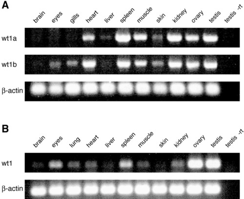
A,B: Expression of wt1 in adult tissues. Indicated organs were isolated from 6 month old zebrafish (A) and mice (B). Wt1 expression levels were determined by RT-PCR. Fragment sizes for zebrafish wt1 were identical to those in Figure 3. In case of the mouse tissues, the wt1 amplicon comprised 194 bp and the β-actin amplicon 165 bp. To verify the absence of genomic DNA contamination, an aliquot of the testis RNA sample from zebrafish and mouse was used for polymerase chain reaction without reverse transcription (-rt). EXPRESSION / LABELING:
|