
- Title
-
Robo3 isoforms have distinct roles during zebrafish development
- Authors
- Challa, A.K., McWhorter, M.L., Wang, C., Seeger, M.A., and Beattie, C.E.
- Source
- Full text @ Mech. Dev.
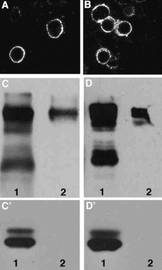
Protein localization of Robo3 isoforms in transiently transfected mammalian cells. myc-tagged (A) robo3var1 and (B) robo3var2 isoforms driven by a CMV promoter were transfected into 293T cells. Protein expression was detected by immunofluorescence using a rhodamine conjugated anti-Myc antibody. (C) 293T cells were transfected with myc-tagged robo3var1 or (D) robo3var2 followed by biotinylation of surface proteins. Lane 1 (C–D′) is the flowthrough and reflects cytoplasmic proteins or proteins that did not bind to avidin. Lane 2 (C–D′) is surface proteins that are biotinylated and bound to avidin. Proteins in C, D are recognized by an anti-myc antibody. (C′, D′) The same blot was stripped and reprobed with anti-actin antibody. Due to lower expression of Robo3var2 in transfected cells, Lanes D 1,2 were exposed longer compared to lanes C 1,2. Lanes C′ and D′ were exposed for the same amount of time reflecting similar amounts of total protein. |
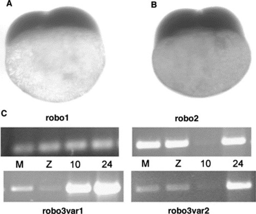
robo orthologs are maternally expressed. Whole-mount RNA in situ hybridization on two-cell stage embryos for (A) robo3var1 and (B) robo3var2 showing maternal expression. RT-PCR experiments using total RNA extracted from different developmental stages: M, maternal (2–512 cell stage); Z, early zygotic (1000 cell stage); 10, 10 hpf and 24, 24 hpf. robo1, robo2, robo3var1 and robo3var2 are expressed at all stages. The expression levels, as indicated by PCR amplifications, are relatively uniform for robo1, decreased expression at 10 hpf for robo2 and robo3var2, and decreased expression at early zygotic stage for robo3var1. Since these primers flank introns, they will not amplify on genomic DNA. |
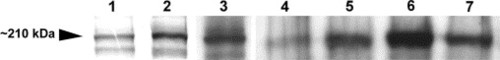
Robo3 protein expression. In vitro translated, Myc-epitope tagged Robo3var1 and Robo3var2 proteins were identified using an anti-Myc antibody (Lanes 1 and 2, respectively). Robo3var2-Myc (Lane 3) and Robo3var1-Myc (not shown) were also recognized by anti-Robo3 antiserum. Robo3 proteins from zebrafish embryo extracts, corresponding to the same size as that of in vitro translated proteins, were also identified by anti-Robo3 antiserum (Lanes 4–7). Robo3 proteins were observed as early as 100% epiboly (Lane 4) and continued to be expressed at 11, 12, and 16–18 hpf (Lanes 5–7, respectively). EXPRESSION / LABELING:
|
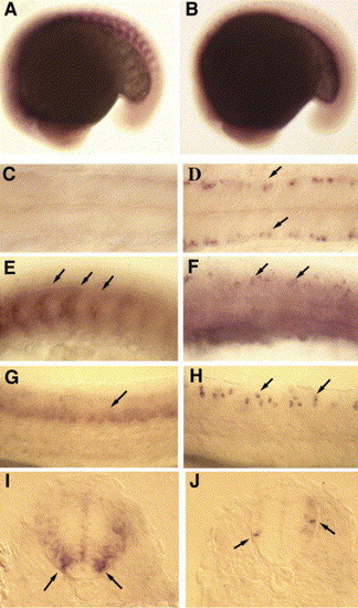
RNA expression patterns of robo3var1 and robo3var2 during the first day of development. Whole-mount RNA in situ hybridization for robo3var1 (A, C, E, G, I) and robo3var2 (B, D, F, H, J). robo3var1 RNA is expressed in trunk somites (A; ∼17 hpf), and at higher magnification (E, arrows) can be seen in the posterior aspect of the somites between 14 and 24 hpf. In contrast, robo3var2 is not expressed in the somites (B; ∼17 hpf) but is seen in the hindbrain (D, arrows, compare to C (dorsal views); ∼17 hpf) and spinal cord (F, arrows; ∼17 hpf) between 14 and 24 hpf. At 24 hpf, robo3var1 is expressed diffusely in the ventral spinal cord (G, arrows), whereas robo3var2 expression is more distinct (H, arrows). Cross-sections of the trunk at 26 hpf reveals that robo3var1 is expressed in ventral and intermediate spinal cord cells (I, arrows), whereas robo3var2 is expressed in dorsal and intermediate spinal cord cells (J, arrows). Anterior is to the left in A–H and dorsal is to the top in I and J. EXPRESSION / LABELING:
|

RNA expression patterns of robo3 isoforms during the second day of development. Lateral (A, C) and dorsal (B, D) views of the head showing whole-mount RNA in situ hybridizations for robo3var1 and robo3var2 at 48 hpf. (A, B) Diffuse expression of robo3var1 in the midbrain (double asterisks in A) with stronger expression in the cerebellum (B, arrows) and hindbrain (B, bar). (C, D) Distinct and robust expression of robo3var2 in the cerebellum (D, arrows) and hindbrain (D, bar). Notice the lack of expression of both isoforms in the tectum (A and C, asterisks). Patches of robo3var2 expressing cells are present in the diencephalic regions (C, arrowhead) and in the telencephalon (D, arrowheads). EXPRESSION / LABELING:
|
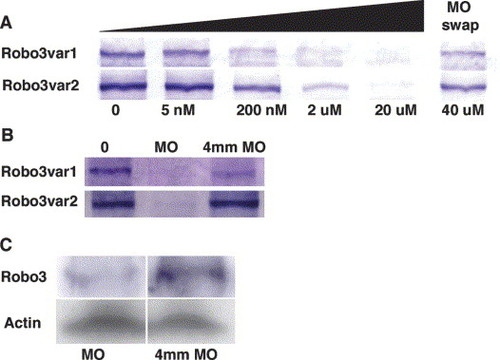
Analysis of robo3var1 and robo3var2 MO efficacy and specificity. Western Blot analysis using anti-Myc (A, B) and anti-Robo3 (C) antibodies. (A) Coupled in vitro transcription–translation in rabbit reticulocyte lysates, using myc-tagged robo3 constructs as templates, in the absence of the corresponding MO or increasing concentrations of the corresponding MO from 5 nM to 20 μM. Forty-micromolar robo3var2 specific MO was added to robo3var1 reaction mixture (MO swap, top row) and vice versa (MO swap, bottom row). (B) Coupled in vitro transcription–translation reactions for Robo3var1 and Robo3var2 in the absence of the corresponding MO, 20 μM of the corresponding MO and 20 μM of the corresponding 4 mm control MO. (C) Total protein extracted from 11 hpf embryos injected with either a mixture of robo3var1 and robo3var2 MOs (MO) or a mixture of robo3var1 4 mm and robo3var2 4 mm control MOs (4 mm MO). Anti-actin antibody (Actin) was used as a protein loading control. EXPRESSION / LABELING:
|
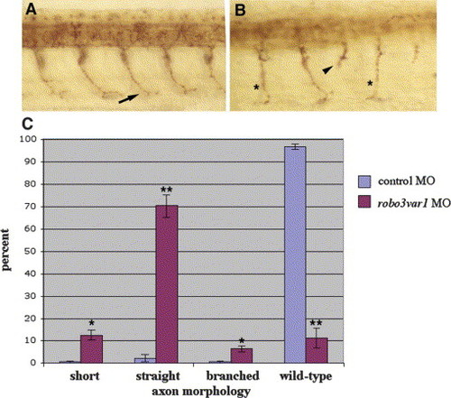
Knockdown of Robo3var1 causes motor axon defects. Lateral views of (A) robo3var1 4 mm control MO and (B) robo3var1 MO injected embryos at 28 hpf labeled with znp1 antibody showing ventrally projecting CaP motor axons. Over 95% of control axons had the stereotyped curved axon morphology and reached the ventral aspect of the myotome in control embryos (A, arrow), whereas straight axons (B, asterisks) and short axons (B, arrowhead) were observed in robo3var1 MO injected embryos. (C) These data were quantitated in control MO (n=103 embryos, 1537 axons, in two experiments) and robo3var1 MO (n=228 embryos, 3527 axons, in three experiments) embryos. *P<0.05; **P<0.001. |
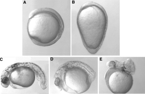
Knockdown of Robo3var2 MO causes morphological defects. Lateral view of live 11 hpf control (A) and (B) robo3var2 MO injected embryos (9 ng). Injecting 6–9 ng of robo3var2 MO resulted in three classes of phenotypes based on severity as visualized at 26–28 hpf. (C) Mildly affected embryos exhibited curved tails, (D) moderately affected embryos had reduced trunk tissue with tail/posterior trunk within the yolk and (E) severely affected embryos had reduced trunk tissues aggregated on the yolk. Seventy percent of the MO injected embryos showed severe to moderate phenotypes (n=300). |

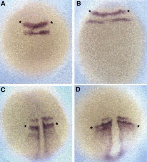
Knockdown of Robo3var2 dorsalizes embryos. Embryos were injected at the 1–2 cell stage with control or robo3var2 MO and processed for RNA in situ hybridization. Embryos were fixed at 70% epiboly and processed for ntl (A, B) and gsc (C, D) in 4 mm control (A, C) and robo3var2 (B, D) MO injected embryos. Embryos were fixed at 14 hpf and processed for scl (E, F) and pax2 (G, H; asterisks denote head expression) in control (E, G) and robo3var2 (F, H) MO injected embryos. Data for gsc (I) and (J) scl were quantitated. Twenty to 70 embryos were scored from at least two separate injection experiments. EXPRESSION / LABELING:
|
Knockdown of Robo3var2 causes expansion of krox20 and papc expression domains. Dorsal views (anterior to the top) of krox20 and papc RNA in situ hybridization at 11 hpf. Six to 9 ng of robo3var2 4 mm control or robo3var2 MO was injected into each embryo. Control embryos did not exhibit any significant defects in the width (asterisks) of krox20 or papc domains (A, C) but robo3var2 MO injected embryos exhibited expansion of both of these expression domains (B, D). EXPRESSION / LABELING:
|

Unillustrated author statements EXPRESSION / LABELING:
|
Reprinted from Mechanisms of Development, 122(10), Challa, A.K., McWhorter, M.L., Wang, C., Seeger, M.A., and Beattie, C.E., Robo3 isoforms have distinct roles during zebrafish development, 1073-1086, Copyright (2005) with permission from Elsevier. Full text @ Mech. Dev.