
- Title
-
Exocrine pancreas development in zebrafish
- Authors
- Yee, N.S., Lorent, K., and Pack, M.
- Source
- Full text @ Dev. Biol.

Adult exocrine pancreas. (A) Whole mount image of the dissected adult zebrafish digestive tract, right lateral view, processed for amylase immunohistochemistry (IHC). Exocrine tissue (green) is identified between adjacent intestinal loops. (B) Adult zebrafish pancreas dissected from adherent intestinal tissue. This fixed specimen reveals the branched network of exocrine lobules and associated ducts. Arrowhead points to insertion site of the extrapancreatic duct to the intestine. An exocrine lobule is outlined by the dashed white line. (C) Histological section of adult pancreas processed for cytokeratin (red) and amylase (green) IHC; DNA counterstained with dapi (blue) to visualize cell nuclei. Dashed white lines outline two acinar glands. Arrows point to large pancreatic ducts within the lobule. Arrowheads point to intercalated ducts. (D) Sagittal histological section showing large pancreatic ducts with adjacent exocrine and intestinal tissues. (E) Histological section of an exocrine lobule showing acinar cells with a prominent duct. Insets (* in panel D; long arrow in panel E) show cuboidal cells lining these ducts. Red dashed line outlines an acinus. (F) Transmission electron micrograph showing an acinus comprised of polarized cells with apical zymogen granules, basal nucleus, mitochondria and rough endoplasmic reticulum. A centroacinar cell is identified based upon its position within the acinar lumen and its scant cytoplasm (inset). e: exocrine tissue; in: intestine; amy: amylase; s: spleen; ag: acinar glands; pd: pancreatic ducts; ic: intercalated ducts; bv: blood vessel; a: acinus; zg: zymogen granules; m: mitochondria; rer: rough endoplasmic reticulum; ca: centroacinar cell; A: anterior; P: posterior; D: dorsal; V: ventral. |
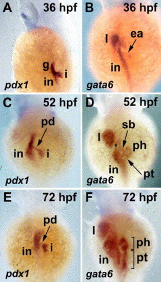
pdx1 and gata6 expression in the developing zebrafish pancreas and digestive tract; whole mount RNA in situ hybridization, dorsal view. (A, B) At 36 hpf, pdx1 expression in the pancreatic islet, intestine and rostral gut endoderm is evident; gata6 is expressed in the intestine, liver and in the exocrine anlage. (C, D) At 52 hpf, pdx1 is expressed in cells of the endocrine pancreas and a stalk of tissue corresponding to the extrapancreatic duct. Histological sections (not shown) show exocrine cells adjacent to the islet also express pdx1. gata6 is expressed in exocrine cells surrounding the islet that forms the pancreatic head. Tissue of the pancreatic tail is first visible at this stage. (*) pancreatic duct insertion site. (E, F) By 72 hpf, the exocrine pancreas has grown significantly; pdx1 expression in the pancreatic duct is diminished. i: pancreatic islet; in: intestine; g: gut endoderm; l: liver; ea: exocrine anlage; pd: pancreatic duct; sb: swim bladder; ph: pancreatic head; pt: pancreatic tail. EXPRESSION / LABELING:
|
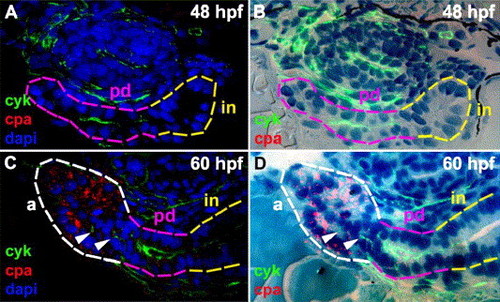
Exocrine differentiation and gland morphogenesis. (A–D) Histological cross-sections of 48 hpf (A) and 60 hpf (C) larvae processed for whole mount cpa and cyk immunohistochemistry (IHC) and dapi. (B and D) Merged fluorescent images without dapi staining (A and C) and bright field images of the corresponding histological sections stained with methylene blue and azure II. The main pancreatic duct (pink outline) is contiguous with the intestine (in, yellow outline) at 48 hpf. Cpa expression is evident in exocrine cells forming a primitive acinus-like structure at 60 hpf (white outline). Cyk+ duct cells (arrowheads) are present adjacent to the developing acinus but are not contiguous with the extrapancreatic duct in this and adjacent sections (not shown). cpa: carboxypeptidase A; cyk: cytokeratin; pd: pancreatic duct; a: acinus; in: intestine. |
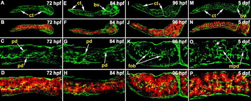
Pancreatic ductal morphogenesis during larval development. (A–P) Confocal images (right, lateral view) of whole mount specimens processed for cyk (green) and cpa (red) immunohistochemistry. (A–D) At 72 hpf, small primary pancreatic ducts are evident, and there is strong cpa staining in acinar cells. (E–H) The 84 hpf pancreas has a few more ducts than at earlier stages. (I–L) At 96 hpf, first order ductal branches are evident. These small ducts appear to extend into the nearby acinar glands. (M–P) At 120 hpf (5 dpf), second order ductal branches (double arrows) that clearly extend into well formed acini are evident. First order ductal branches (single arrow) and main pancreatic duct segments are also evident at this stage. cyk: cytokeratin; cpa: carboxypeptidase A; pd: pancreatic ducts; fob: first order ductal branches; mpd: main pancreatic duct; ct: connective tissue; bv: blood vessel. EXPRESSION / LABELING:
|
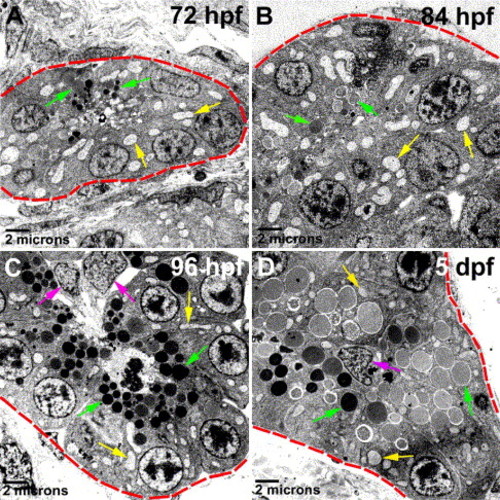
Acinar cell ultrastructure. (A–D) Transmission electron micrographs of pancreatic acini (outlined by red dashed lines). Progressive maturation of acinar cells is evident between 72 hpf and 5 dpf as determined by the cell polarity, number and size of zymogen granules (green arrows) and the amount of mitochondria (yellow arrows) and rough endoplasmic reticulum. Centroacinar cells are indicated by pink arrows. |
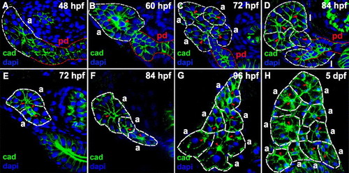
Acinar gland morphogenesis during larval development. (A–D) Histological sections through the anterior pancreas (rostral to islet) of larvae processed for cadherin (green) immunohistochemistry and dapi (blue). The main pancreatic duct and the acini are demarcated (red and white dashed lines respectively), and centroacinar cells are indicated (red arrowheads). (A) At 48 hpf, the distal end of the main pancreatic duct appears as a multi-layered stratified epithelium (white dashed lines). (B) By 60 hpf, a primitive acinus-like structure is formed. (C) At 72 hpf, four distinct acini are recognized. (D) By 84 hpf, two exocrine lobules are evident. (E–H) Histological cross-sections through the pancreas (caudal to islet) of larvae processed for cadherin immunohistochemistry (green) and dapi (blue). Acini (demarcated by white dashed lines) are shown. Acinar number increases on successive days post-fertilization. Two, three, six and nine acini are identified in these 72 hpf (E), 84 hpf (F), 96 hpf (G) and 5 dpf (120 hpf) (H) larvae, respectively. pd: pancreatic duct; a: acinus; l: exocrine lobule; cad: cadherin. |
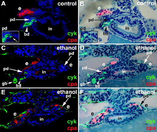
Ethanol treatment disrupts exocrine pancreas morphogenesis. Histological cross-sections (viewing posteriorly) of a 4 dpf control (A, B) and an ethanol-treated larva (C–F) processed for cyk (green) and cpa (red) immunohistochemistry. (A, C, E) Fluorescent images with dapi (blue); (B, D, F) merged fluorescent images without dapi and bright field images of the corresponding histological sections stained with methylene blue and azure II. (A, B) The extrapancreatic duct and extra-hepatic bile duct of control larvae join the intestine at the same level. Inset shows site of pancreatic duct insertion into the intestine (arrow). (C–F) Bilateral cyk+ and cpa+ exocrine tissues are present in ethanol-treated larva. Bilateral extrapancreatic ducts that fail to join the intestine in these sections, as well as in consecutive rostral and caudal sections (not shown). Sections depicted in panels (C), (D) and (E), (F) are separated by 9 μm (C and D rostral to E and F). cyk: cytokeratin; cpa: carboxypeptidase A; pd: pancreatic duct; bd: bile duct; in: intestine; e: exocrine tissues; gb: gall bladder. EXPRESSION / LABELING:
|

Mutations affecting early stages of exocrine pancreas development on 4 dpf. Right lateral view of live wild type (A), slj (B), pie (C) and flo (D) larvae. In the wild type larva (A), the intestinal epithelium is folded, only a small amount of yolk is evident, and the swim bladder is inflated. By comparison, the intestines in slj (B), pie (C) and flo (D) are narrow, and they lack folds. (E–H) Histological cross-sections through the exocrine pancreas 3 μm caudal to the islet. Wild type acinar cells contain zymogen granules (E, red arrows), whereas in slj (F), pie (G) and flo (H), the pancreatic cells lack zymogen granules. (I–L) Dorsal view of larvae processed for whole mount RNA in situ hybridization using an antisense trypsin probe. Strong trypsin expression is present in the wild type larva (I), whereas in slj (J), pie (K) and flo (L) larvae, there is reduced trypsin expression. Arrows point at pancreatic head and pancreatic tail. (M–P) Wild type and mutant pancreata processed for whole mount cyk and cpa immunohistochemistry (IHC). The wild type exocrine pancreas (M) is comprised of cpa+ acinar cells and a network of cyk+ ducts that joins the extrapancreatic main duct. The exocrine pancreata of slj (N), pie (O) and flo (P) larvae are small. The extrapancreatic duct can be identified, but there is no significant cpa detected, and only a small number of ducts are recognizable. (Q–T) Confocal analysis of pancreatic ducts following cyk IHC. Ducts in the mutant pancreata resemble primary ducts seen in wild type larvae (Figs. 4C, G). (U–X) Histological cross-sections through the pancreas ∼3 μm posterior to the islet of larvae processed for cyk IHC merged with bright field images of the corresponding histological sections stained with methylene blue and azure II. Small ducts are evident in the wild type larva (U; arrowheads) and in slj (V), pie (W) and flo (X) larvae). slj: slimjim; pie: piebald; flo: flotte lotte; e: exocrine pancreas; in: intestine; cyk: cytokeratin; cpa: carboxypeptidase A; epd: extrapancreatic main duct. EXPRESSION / LABELING:
|

Mutations affecting late stages of exocrine pancreas development. (A1–E1) Right lateral view of live 5 dpf wild type and mutant larvae. (A1) The wild type intestinal epithelium is folded, a widely patent gut lumen is obvious, and there is little residual yolk. The intestine is grossly normal in swd (B1), mms (C1), djm (D1) and dtp (E1) mutants. In swd, skin pigmentation (melanophores) is reduced, but xanthophores and iridophores appear unaffected (B1). There is a small amount of residual yolk in dtp (E1). (A2–E2) Histological cross-sections through the 5 dpf pancreas, 3 μm caudal to the islet. In wild type (A2), there are multiple exocrine acini; in swd (B2), there are relatively few exocrine acini, and electron micrograph shows that the acinar cells contain zymogen granules (Supplementary Fig. 3D); in mms (C2), multiple acini are observed, but the cytoplasm of the acinar cells contains vacuole-like structures and very few zymogen granules (Supplementary Fig. 3H); in djm (D2) and dtp (E2), there are no recognizable acini present, and the exocrine cells appear undifferentiated. (A3–E3) Right lateral view of whole mount trypsin in situ hybridization on 5 dpf. Strong trypsin expression is evident in the wild type pancreas (A3), whereas in swd (B3), mms (C3), djm (D3) and dtp (E3), trypsin expression is reduced. There is background staining in the yolk. (A4–E4) Visualization of pancreatic ducts and acinar cells by cyk and cpa immunohistochemistry, respectively. The wild type ductal system is composed of a branching network of small ducts and cpa+ acinar cells (A4). The swd pancreas (B4) is small, but there is relatively normal cpa; in the mms pancreas (C4), there are only low levels of cpa. There is no significant cpa detected in djm (D4) and dtp (E4). (A5–E5 and A6–E6) Confocal projection of pancreatic ducts (green) and acini (red) in larvae processed for cyk and cpa immunohistochemistry. Wild type pancreatic ducts are highly branched, and individual acini are each drained by single intercalated duct (A5, 6). In swd (B5, 6), the main pancreatic duct with few branches is evident, whereas in mms (C5, 6), duct branching is evident, albeit less pronounced than in wild type. By comparison, the ductal networks of djm (D5, 6) and dtp (E5, 6) are markedly aberrant. swd: sweetbread; mms: mitomess; djm: ductjam; dtp: ducttrip; e: exocrine pancreas; in: intestine; y: yolk; cyk: cytokeratin; cpa: carboxypeptidase A. EXPRESSION / LABELING:
|
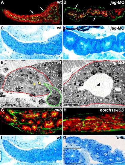
Notch signaling regulates pancreatic duct development. (A, B) Sagittal optical section (A) and histological section (B) of the pancreas of wild type (A) and jagged2/3 morpholino-injected (B) 5 dpf larvae processed for cpa (red) and cyk (green) immunohistochemistry (IHC), right lateral view. Cpa is present in the jagged morpholino-injected larva (B), although the levels are reduced compared with wild type (A). By contrast, the ductal system is markedly reduced in the jagged morpholino-injected larva (B). (C, D) Sagittal histological sections through the pancreas of 5 dpf wild type (C) and jagged morpholino-injected (D) larvae. Note the contiguous enlarged acini present in the jagged morpholino-injected larva. By contrast, individual wild type acini are difficult to delineate because they are numerous, relatively small and in close proximity to each other. (E, F) Transmission electron micrographs showing acini (red dashed lines) from wild type (E) and jagged morpholino-injected (F) larvae. The acinar lumen of the jagged morpholino-injected larva is dilated, and centroacinar cells are absent. An intercalated duct exiting the wild type acinus is evident (green dashed line), and two centroacinar cells are indicated by yellow arrowheads. (G, H) Confocal projection of larvae processed for cyk (green) and cpa (red) IHC. Cyk IHC shows altered ductal morphology in mib (G) and Notch-activated (H) larvae. Cpa IHC shows clusters of enlarged acini in mib (G), whereas following Notch activation, the level and distribution of cpa protein are near normal (H), compared with wild type pancreas (Figs. 4O and P; Figs. 9A5 and A6). (I, J) Sagittal histological sections through the 5 dpf wild type (I) and mib (J) pancreas, right lateral view. Note enlarged acini (inset showing a magnified view) in the mib larva. jag-MO: jagged2/3 morphant; mib: mind bomb; notch1a-ICD: notch1a-intracellular domain; cpa: carboxypeptidase A; cyk: cytokeratin; al: acinar lumen. Arrows in panels (A) and (B) point to immunoreactive cyk in pancreatic connective tissue. EXPRESSION / LABELING:
|
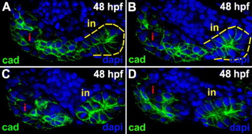
Exocrine anlage architecture. (A–D) Serial 3 μm sections through the pancreas of a 48 hpf zebrafish larva processed for cadherin (green) immunohistochemistry and dapi (blue). Exocrine cells surround the islet, and they are adjacent to the intestine (yellow dashed line). These cadherin immunostainings show that the exocrine cells are not arranged as a simple columnar epithelium. Compare appearance of the intestinal epithelium, which in these sections is cut tangentially, to the exocrine cells. Most intestinal epithelial cells have a columnar appearance, whereas the exocrine cells appear round or have a stellate appearance and they are stratified. cad: cadherin; i: islet; in: intestine. |

Trypsin expression by antisense RNA in situ hybridization in exocrine pancreas mutants at 72 hpf. Compared with wild type larva (A), there are reduced numbers of trypsin+ exocrine cells in slj (B), pie (C) and flo (D) larvae. Reduced trypsin expression is also evident in dtp (E), but exocrine size is normal. The 72 hpf dtp larvae identified on the basis of altered trypsin expression in 25% of progeny of heterozgyous dtp fish. slj: slimjim; pie: piebald; flo: flotte lotte; dtp: ducttrip. Note: Trypsin expression in the other 72 hpf mutants (swd, mms and djm) is indistinguishable from wild type (data not shown). EXPRESSION / LABELING:
|

Exocrine pancreas in the sweetbread (swd) and mitomess (mms) mutants. (A, B) Right lateral view of 72 hpf wild type (A) and swd (B) larvae processed for trypsin whole mount in situ hybridization. There is normal trypsin expression in the 72 hpf swd mutant identified by reduced skin pigmentation (B). (C, D) Acinar cell hypoplasia is evident in 5 dpf swd mutant. Acinar cells (red dashed lines) in swd mutant (D) appear small with relatively few and small zymogen granules (indicated by green arrows), compared with wt sibling (C). (E, F) Histological cross-sections through the pancreatic tail of 5 dpf wild type (E) and mms (F) mutant larvae stained with methylene blue and azure II. In routine histological sections, acinar gland morphology is difficult to appreciate. However, both in wild type and mms mutants, acinar cells surrounding centroacinar cells (yellow arrowheads) can be discerned. Red arrow heads outline acinar borders. Note the zymogen granules are prominent in wild type and they are relatively few and small in mms. Also note the cytoplasmic vacuole-like structures in mms acinar cells. Electron microscopy identifies these as swollen mitochondria (below). Inset (a) shows preserved mms acinar gland morphology as revealed by cadherin (green) immunohistochemistry and dapi (blue). (G, H) Transmission electron micrographs of 5 dpf wild type and mms acinar cells. Note reduced numbers and size of mms acinar zymogen granules and enlarged swollen mitochondria. zg: zymogen granules; m: mitochondria; a: acinus; cad: cadherin. EXPRESSION / LABELING:
|

Unillustrated author statements EXPRESSION / LABELING:
|
Reprinted from Developmental Biology, 284(1), Yee, N.S., Lorent, K., and Pack, M., Exocrine pancreas development in zebrafish, 84-101, Copyright (2005) with permission from Elsevier. Full text @ Dev. Biol.