
- Title
-
A pair of Sox: distinct and overlapping functions of zebrafish sox9 co-orthologs in craniofacial and pectoral fin development
- Authors
- Yan, Y.L., Willoughby, J., Liu, D., Crump, J.G., Wilson, C., Miller, C.T., Singer, A., Kimmel, C., Westerfield, M., and Postlethwait, J.H.
- Source
- Full text @ Development

Embryonic expression patterns. A,C,D,G,I,K,L, sox9a; B,E,F,H,J,M,N, sox9b. (A,B) 14 hpf, dorsal view. (C,F) Cross-sections of 14 hpf embryos at anterior-posterior level indicated by black bars in A and B. (D,E) Lateral views of 26 hpf embryos. (G,H) Lateral view, 48 hpf. (I,J) Pectoral fin bud at 68 hpf. (K-N) High (K,N) and low (L,M) magnification of sections of 68 hpf embryos through the arches. Abbreviations: co, scapulocoracoid; f, forebrain; fb, fin bud; e, eye; ed, endochondral disc; hb, hindbrain; m, midbrain; mhb, midbrain-hindbrain boundary; nc, neural crest; ov, otic vesicle; pa, pharyngeal arches; pcp, prechordal plate; r, rhombomeres; s, somites; t, tectum. Scale bars: in N, 50 µm for K,N; in B, 100 µm for A,B; in F, 100 µm for C-F; in H, 100 µm for G,H; in J, 100 µm for I,J; in M, 100 µm for L,M; in N, 100 µm for K,N. EXPRESSION / LABELING:
|

sox9bb971 linkage map and sox9b expression, and sox9b morpholino-treated embryos. (A) LG3 (distance in cM), with red markers shown to be deleted, blue markers shown not to be deleted, and black markers untested. Region deleted, pale red. (B,C) Lateral views of 24 hpf embryos probed for sox9b expression. (B) wild type; (C) sox9bb971. Mutant embryos lack sox9b expression. (D,E) Dorsal, right side view of cranial crest in four-somite stage embryos probed for sox9b expression. (D) Wild type; (E) embryo injected with sox9b splice-directed MO showing retention of sox9b transcript in the nuclei. Scale bars: in C, 100 µm for B,C; in E, 100 µm for D,E. EXPRESSION / LABELING:
|

Wild-type, sox9 mutants, and sox9b-morpholino-treated animals. (A-E) Live larvae; (F-J) lateral view of head of Alcian-stained larvae. (K-O) Flat mount of Alcian-stained pharyngeal arches. (P-T) Flat mount of Alcian-stained neurocranium. (U-Y) Alcian-stained pectoral fin bud. All animals at 4 dpf. (A,F,K,P,U) Wild type; (B,G,L,Q,V) sox9a mutant, showing deletion of most cranial cartilage and scapulocoracoid; (C,H,M,R,W) sox9bb971; neurocranium, arches, and endochondral disc reduced; (D,I,N,S,X) sox9b MO-injected animals phenotypically similar to sox9bb971; (E,J,O,T,Y) double mutant, arches, neurocranium, and fin deleted. Abbreviations: abc, anterior basicranial commissure; cbs, ceratobranchials; ch, ceratohyal, m, Meckel's cartilage; cl, cleithrum; co, scapulocoracoid; ed, endochondral disc; ep, ethmoid plate; hs, hyosymplectic; j, jaw; ov, otic vesicles; no, notochord; pc, parachordal; pq, palatoquadrate; tr, trabecula. Scale bars: in E, 100 µm for A-E; in T, 100 µm for F-T; in Y, 100 µm for U-Y. PHENOTYPE:
|

Appendage developmental control genes in sox9 mutants. (A-H) The apical ectodermal ridge gene dlx2a. (I-P) The zone of polarizing activity gene shh. (A-D,I-L) 34 hpf; (E-H,M-P) 52 hpf. (A,E,I,M) Wild type; (B,F,J,N) sox9a-; (C,G,K,O) sox9bb971; (D,H,L,P) sox9a-; sox9bb971 double mutant. Scale bar: in P, 100 µm. |

Injection of sox9b mRNA partially rescues the mutant phenotype. (A,B) Wild type; (C,D) Control sox9bb971; (E-J) sox9b mRNA-injected sox9bb971 larvae, showing partial rescue of the craniofacial skeletal phenotype of sox9bb971. Partial rescue occurred in 14 of 85 injected four-day-old homozygous mutant larvae. (A,C,E,G,I) Pharyngeal arches; (B,D,F,H,J) Neurocranium. Scale bar: in J, 100 µm. |

Bone development. (A-E) Lateral view of runx2b expression at 2 dpf. (F-J) Alizarin red staining on live larvae at 5 dpf. (A,F) Wild type; (B,G) sox9a-; (C,H) sox9bb971; (D,I) sox9b MO injected; (E,J) double mutant. Abbreviations: I, first pharyngeal arch; II, second pharyngeal arch; bsr, brachiostegal rays; cb5, ceratobranchial arch 5; ch, ceratohyal; cl, cleithrum; de, dentary; ent, entopterygoid; hm, hyomandibular; max, maxilla; op, opercle; ot, otolith; ps, parasphenoid; ra, retroarticular. Scale bar: in J, 100 µm. |
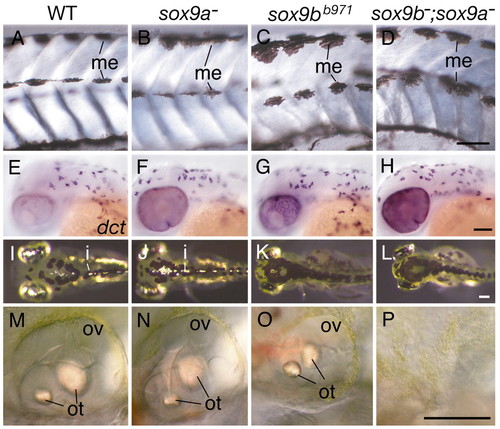
Pigment cells and ears in mutants and wild types. (A-D) Trunk melanocytes, lateral view at 4 dpf, showing large melanocytes in sox9bb971 and double mutant. (E-H) dct expression at 26 hpf, showing little difference among genotypes; (I-L) iridophores at 4 dpf, showing fewer iridophores in sox9bb971 and double mutant. (M-P) ear at 4 dpf, showing small ear in sox9bb971 and no ear in double mutant. (A,E,I,M) Wild type; (B,F,J,N) sox9a- mutant; (C,G,K,O) sox9bb971; (D,H,L,P) double mutant. Abbreviations: i, iridophore; me, melanocyte; ot, otolith; ov, otic vesicle. Scale bars: in D 100 µm for A-D; in H, 100 µm for E-H; in L, 100 µm for I-L; in P, 100 µm for M-P. |
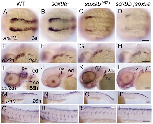
Expression of markers for neural crest and collagen in wild-type and mutant embryos. (A-D) Dorsal view of snail1b expression at three-somite stage showing decreased expression in sox9bb971 and double mutant; (E-T) lateral views; (E-H) dlx2a expression at 24 hpf slightly reduced in sox9bb971 and double mutant; (I-L) col2a1 expression at 68 hpf showing lack of scapulocoracoid in sox9a mutant and double mutant and smaller endochondral disc in sox9bb971 and double mutant; (M-T) sox10 expression in trunk and tail of 26 hpf embryos showing decreased expression. (A,E,I,M) Wild type; (B,F,J,N,R) sox9a- mutant; (C,G,K,O,S) sox9bb971; (D,H,L,P,T) double mutant. (Q-T) Higher magnification of M-P. Abbreviations: co, scapulocoracoid; ed, endochondral disc; ov, otic vesicle. Scale bars: in D 100 µm for A-D; in H, 100 µm for E-H; in L, 100 µm for I-L; in P, 100 µm for M-P; in T, 100 µm for Q-T. EXPRESSION / LABELING:
|

Ectopic expression of sox9a and sox9b mRNAs causes ectopic expression of other neural crest genes. (A,D,G,J,M) Uninjected controls. (B,E,H,K,N) Embryos injected with sox9a mRNA. (C,F,I,L,O) Embryos injected with sox9b mRNA. (A-C) Embryos probed for sox9a. (D-F) sox9b probe; (G-I) foxd3 probe; (J-L) sox10 probe; (M-O) snai1b probe; vertical line indicates anterior border of the neural plate. Scale bar: in O, 100 µm. EXPRESSION / LABELING:
|
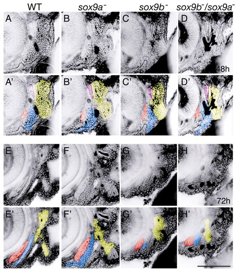
Differential functions of sox9 duplicate genes in the morphogenesis and growth of chondrogenic crest. Confocal micrographs of the pharyngeal arches of wild type (A,A',E,E'), sox9a- (B,B',F,F'), sox9bb971 (C,C',G,G'), and sox9bb971; sox9a-. (D,D',H,H') animals stained with the vital dye, BODIPY-ceramide as described (Yan et al., 2002) to label interstitial spaces. Images are color reversed so cells appear white and interstitial spaces black: original images (A-H) have been psuedocolored (A'-H') to highlight arch features. (A-D') At 48 hours, prechondrogenic condensations of crest cells are seen in the first (blue) and second (yellow) arches. The adductor mandibulae muscle core (red) and first endodermal pouch (purple) are shown for reference. Prechondrogenic condensations are normal in both single mutants (B-C') and reduced in size in the double mutant (D,D'). (E-H') The same individual animals in A-D ' were imaged at 72 hours. In wild-type animals (E,E'), chondrocytes have arranged themselves into characteristic stacks and are easily visualized by their thicker cell matrix. First arch cartilages include Meckel's (m) and palatoquadrate (pq), and second arch cartilages include the hyosymplectic (hs) and ceratohyal (ch). As described in Yan et al. (Yan et al., 2002), in sox9a- animals, crest condensations are present but fail to differentiate and undergo morphogenesis to form stacks of chondroctyes (F,F'). In contrast, in sox9bb971 animals (G,G'), chondrocytes are greatly reduced in number, yet can still form small stacks (e.g. pq). The sox9bb971; sox9a- double mutants show an additive effect including both morphogenetic and growth defects: condensations are smaller in size and do not form stacks of chondrocytes. Total animals examined: nwt=3, nsox9a=1, nsox9b=4, nsox9a;sox9b=5. Scale bar: in I', 100 µm. PHENOTYPE:
|
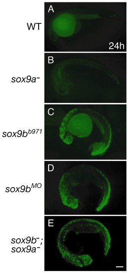
Cell death in wild-type and mutant embryos. Lateral views of TUNEL-labeled embryos at 24 hours. (A) wild type; (B) homozygous sox9a- embryo; (C) homozygous sox9bb971mutant embryo; (D) sox9b-MO-treated wild-type embryos; (E) double mutant. Cell death is increased in sox9bb971 mutants, sox9b MO-treated embryos, and in double mutants. Scale bar: in E, 100 µm. |

Unillustrated author statements PHENOTYPE:
|