- Title
-
Role of SDF1 chemokine in the development of lateral line efferent and facial motor neurons
- Authors
- Sapede, D., Rossel, M., Dambly-Chaudiere, C., and Ghysen, A.
- Source
- Full text @ Proc. Natl. Acad. Sci. USA
|
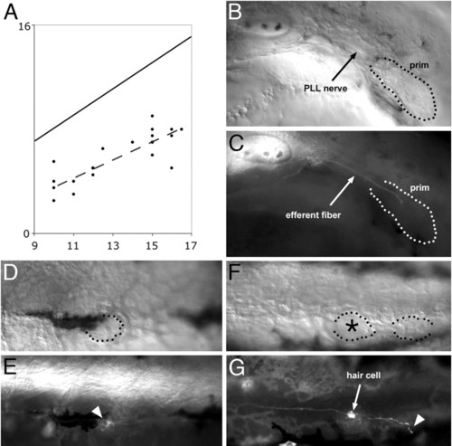
Peripheral growth of the efferent axons. (A) plot of the position of the leading efferent growth cones (ordinate) as a function of the developmental stage of the embryo (abscissa). The developmental stage is expressed as the position reached by the leading edge of the primordium, which is more accurately defined than its trailing edge (15). Both the position reached by the leading efferent neurite (ordinate) and the position reached by the leading edge of the primordium (abscissa) are expressed as somite number. Because the primordium extends over 2.5 somites on average, the diagonal line represents the average position of the trailing edge of the primordium. In this sample of 19 embryos, the leading efferent neurite lags anywhere between 3 and 9 somites behind the trailing edge of the primordium. The dashed line is the least-squares regression line for the positions of the efferent leading edges. (B and C) In a 32 hours after fertilization (haf) sdf1a morphant embryo, where the primordium fails to migrate (B), the efferent fiber reaches the trailing region of the primordium and stops there (C). When the primordium eventually differentiates into one or two ectopic neuromasts, these are invariably innervated by efferent neurites (not shown). (D–G) Efferent innervation in 3-day-old Zath1 morphant embryos. (D and E) A neuromast that comprises no hair cells (dotted outline) is nevertheless contacted by an efferent fiber (arrowhead). (F and G) Two terminal neuromasts, one of which (asterisk) comprises a hair cell and the other not. The neuromast with no hair cell is contacted by an efferent fiber (arrowhead). (Final magnification: B and C, x150; D–G, x130.) |
|
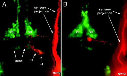
Localization of caudal PLL efferent neurons relative to the facial motor nucleus as visualized in islet-GFP embryos. (A) Two efferent neurons, n1 and n2, occupy normal positions in a 3-day-old larva. GFP labeling is in green, DiI labeling is in red. (B) in a 48-haf embryo, the cell body is located at the posterior edge of the facial nucleus (VII) and has not yet reached its final, more posterior, position. The labeled efferent neurons appear red rather than yellow because the islet-GFP labeling was relatively faint and because the DiI labeling was enhanced to allow visualization of the axon (and partly of the dendrite, dend). gang, Ganglion. |
|
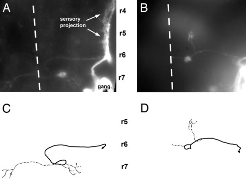
Central pathway of caudal PLL efferent neurons in 3-day-old embryos. (A and C) In the wild type. (B and D) In a sdf1a morphant embryo. A and B have been assembled by using a set of photographs taken at various focal planes; C and D have been drawn on the basis of A and B. Solid lines, axons; dotted lines, dendrites; dashed line, midline; r4, r5, r6, and r7, rhombomeres 4, 5, 6, and 7. In this and in the following figures, the approximate positions of rhombomeres shown on the figures are based on two criteria: the position of the otic vesicle, which forms along rhombomere 5 and extends approximately from rhombomere 4 to rhombomere 6, and the position of identified cranial nerves, specifically the V and VII nerves that exit the brain at the levels of rhombomeres 2 and 4, respectively, and were visualized by fluorescence in islet-GFP embryos, both in wild type and inmorphants. |

Central pathway of rostral and caudal PLL efferents in 3-day-old embryos. (A and C) Wild type. (B and D) sdf1a morphant. Symbols and abbreviations are as in Fig. 3. de, axon of a diencephalic efferent neuron. |
|
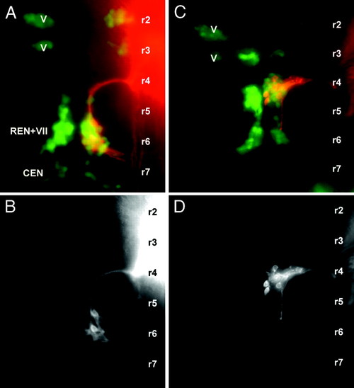
Central pathways of facial motor neurons in 2-day-old wild type (A and B) and sdf1a morphant embryo (C and D). The facial neurons were labeled with DiI and are shown either alone (B and D) or in an islet-GFP background (A and C; red, DiI; green, GFP). The approximate localization of the rhombomeres (r1, r2,...) was defined as in Fig. 3. V, trigeminal motor nucleus; VII, facial motor nucleus; REN, rostral efferent nucleus of the lateral line system; CEN, caudal efferent nucleus of the lateral line system. |
|
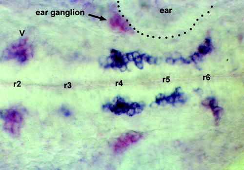
Expression of cxcr4b (blue) and of islet (purple) in a 25-haf embryo. Two streams of doubly labeled cells extend from r4 to r6 on either side of the midline. At the level of r6 the streams turn laterally and begin to loose cxcr4b expression, such that the purple labeling becomes more visible. In a few of the clusters that express islet-1, cxcr4b is expressed at a low level (trigeminal motor neurons in r2, labeled V in the figure) or not at all (statoacoustical ganglion apposed to the ear vesicle, labeled ear ganglion in the figure). EXPRESSION / LABELING:
|

