
- Title
-
The chianti zebrafish mutant provides a model for erythroid-specific disruption of transferrin receptor 1
- Authors
- Wingert, R.A., Brownlie, A., Galloway, J.L., Dooley, K., Fraenkel, P., Axe, J.L., Davidson, A.J., Barut, B., Noriega, L., Sheng, X., Zhou, Y., Tübingen 2000 Screen Consortium, and Zon, L.I.
- Source
- Full text @ Development
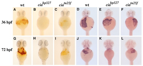
Characterization of the embryonic blood phenotype in cia. (A-L) Ventral views of the anterior region of embryos. (A-C,G-I) Whole-mount o-dianisidine staining of wild-type and cia embryos. Compared with wild type at 36 hpf (A), ciahp327 (B) (shown as representative of ciahs019 and ciaiu089 at all stages) lack hemoglobinized erythrocytes, while ciatu25f (C) manifest a moderate decrease. At 72 hpf, circulating hemoglobinized erythrocytes are still absent in ciahp327 (H) and a moderate decrease is again observed in ciatu25f (I) compared with wild type (G). (D-F,J-L) Whole-mount RNA in-situ hybridization for ße1 globin in wild-type and cia embryos. (D-F) At 36 hpf, cia embryos are indistinguishable from wild type, while the onset of anemia in cia is apparent at 72 hpf, with ciahp327 (K) possessing less than approximately 30% of cells compared with wild type (J), and ciatu25f (L) exhibiting an approximate 50% decrease in erythrocytes. PHENOTYPE:
|

Adult blood characterization in ciatu25f and ciaiu089. (A,B) Wright-Giemsa staining of peripheral blood collected from wild-type zebrafish adults and cia shows that mutant red blood cells are visibly microcytosed, and reveals the presence of undifferentiated cells (arrows) in circulation. (C,D) Kidney samples from wild-type and cia adults shows an increased number of erythroid precursors of cia mutants, as well as markedly increased cellularity. Scale bars: 20 µm in A,B; 40 µm in C,D. PHENOTYPE:
|

Expression patterns of zebrafish tfr1a and tfr1b during embryogenesis. Whole-mount RNA in-situ hybridization for tfr1a (B,E,H) shows an expression pattern restricted to the hematopoietic intermediate cell mass and later circulating blood, identical to that of ße1 globin (A,D,G), shown at 15 somites, 20 hpf, and 36 hpf. By contrast, the expression of tfr1b (C,F,I) at these timepoints is ubiquitous. EXPRESSION / LABELING:
|
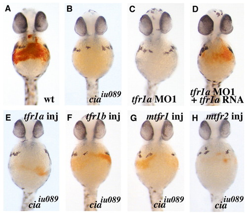
tfr1a is required for erythrocyte hemoglobin production, but multiple Tfr family members can compensate for loss of tfr1a in cia. (A-H) Ventral views of the anterior region of o-dianisidine stained embryos at 40 hpf. (A) Uninjected wild type. (B) Uninjected ciaiu089. (C) Wild-type embryo injected with tfr1a MO1 did not exhibit hemoglobinized erythrocytes. (D) Wild-type embryo co-injected with tfr1a MO1 and tfr1a cRNA was partially rescued. (E-H) ciaiu089 embryos injected with cRNA of tfr1a (C), tfr1b (D), mouse tfr1 (E), and mouse tfr2 (F) all exhibited partial rescue of hypochromia. PHENOTYPE:
|
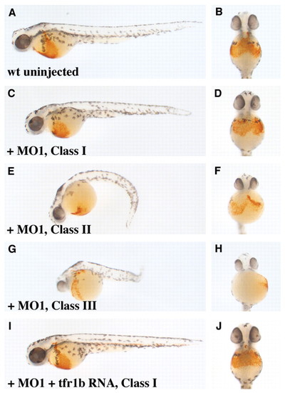
Functional analysis of zebrafish tfr1b using morpholinos. (A,C,E,G,I) All show lateral views of 48 hpf o-dianisidine stained embryos, anterior to the left, with (B,D,F,H,J) showing ventral views of the same embryos. (A) Uninjected wild type. (C-H) Wild-type embryos injected with tfr1b MO1 exhibit three categories of phenotypic classes: (C,D) Class I embryo; (E,F) Class II embryo; and (G,H) Class III embryo. (I,J) Embryo co-injected with tfr1b MO1 and tfr1b cRNA. |

Unillustrated author statements EXPRESSION / LABELING:
PHENOTYPE:
|