
- Title
-
Roles for GFRalpha1 receptors in zebrafish enteric nervous system development
- Authors
- Shepherd, I.T., Pietsch, J., Elworthy, S., Kelsh, R.N., and Raible, D.W.
- Source
- Full text @ Development
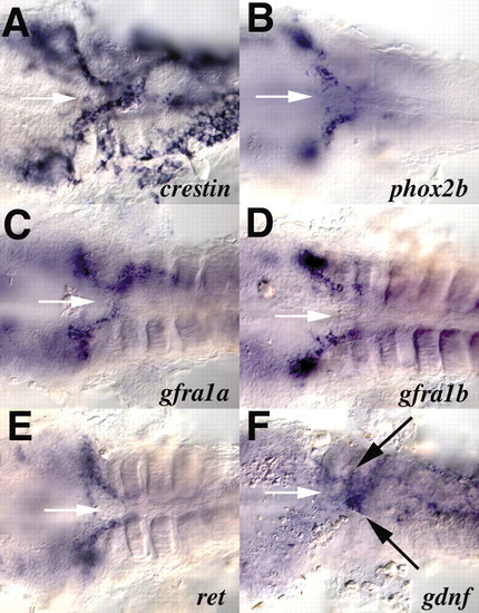
Expression of gdnf receptor components in enteric precursors. Shown are ventral views of the vagal region of 36 hpf embryos after the yolk has been removed. Anterior is towards the left. In situ hybridization was performed for crestin (A), phox2b (B), gfra1a (C), gfra1b (D), ret (E) and gdnf (F). White arrows (A-E) indicate the anterior end of the gut tube. Black arrows in F indicate the mesendodermal expression of gdnf at the anterior end of the gut tube. EXPRESSION / LABELING:
|

Expression of gfra1a, gfra2 and phox2b message in the enteric nervous system. Lateral views of the trunk of 48 hpf embryos, somites 1-11, with the yolk removed (A,B), and the trunk of 96 hpf embryos from somites 10-14 (C,D) that have been hybridized with riboprobes for gfra1a (A,C), phox2b (B) and gfra2 (D). (E) Transverse section taken through a 96 hpf embryo at the level of somite 14 that has been hybridized with probe for gfra1a. Arrows in A,B indicate gene expression in the gut tube. Arrows in E indicate enteric neurons expressing gfra1a message around the circumference of the gut tube. The broken white line in E indicates the border of the mucosa while the broken green line indicates the gut wall. (A-D) Anterior is towards the left. |

Expression of gdnf receptor components in cranial ganglia and brain. Lateral views (A-D) and dorsal views (E-H) of 24 hpf (A,C) and 48 hpf (B,D-H) embryos. At 24 hpf, gfra1a (A) is expressed in lateral line ganglia, while gdnf (C) is expressed in the migrating lateral line primordium (arrow). At 48 hpf, gfra1a (B) is expressed in epibranchial ganglia, while gdnf (D) is expressed in pharyngeal pouches (arrows). gfra1a (E), gfra2 (F), ret (G) and th (H) are shown at 48hpf. Black arrowheads indicate the ventral diencephalic neurons, red arrowheads indicate dorsal diencephalic neurons, white arrowheads indicate the ventral midbrain neurons, pink arrowheads indicate trigeminal motor nucleus, green arrowheads indicate anterior hindbrain domain. all, anterior lateral line ganglia; pll, posterior lateral line ganglia; llp, lateral line primordium; gV, trigeminal ganglia; gVI, facial ganglion; gVIII, octaval/statoacoustic ganglion; gIX, glossopharyngeal; gX, vagal ganglion; DiC, diencephalic catecholaminergic cell cluster; LC, locus coeruleus; sym, sympathetic neurons. EXPRESSION / LABELING:
|

Effect of gfra1a, gfra1b and ret antisense morpholino oligonucleotide injection on enteric neuron development. Shown are lateral views of the guts of 96 hpf embryos stained with anti-Hu antibody, anterior is towards the left. Arrows indicate the end of the gut. (A) Control embryo. (B) 5 ng gfra1a morpholino injected embryo. (C) 5 ng gfra1b morpholino injected embryo. (D) 5 ng gfra1a plus 5 ng gfra1b morpholino injected embryo. (E) 10 ng gfra2 morpholino injected embryo. (F) 10 ng ret morpholino injected embryo. PHENOTYPE:
|

mRNA rescue of morpholino injections. Shown are lateral views of the guts of 96 hpf embryos stained with anti-Hu antibody, anterior is towards the left. Arrows indicate the end of the gut. Embryos were injected with 5 ng gfra1a morpholino (A-C), 5 ng gfra1a plus 5 ng gfra1b morpholino (D,E) or 10 ng ret morpholino (F,G), combined with 50 pg gfra1a* mRNA (A,D), 50 pg gfra1b mRNA(B,F) or 50 pg gfra2 mRNA (C,E,G). |
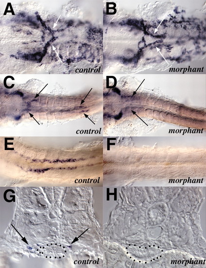
Co-injection of gfra1a and gfra1b morpholinos causes the failure of enteric precursor migration along the gut but does not perturb the initial migration of vagal neural crest to the anterior gut. (A,C,E,G) Control embryos and (B,D,F,H) embryos co-injected with gfra1a plus gfra1b morpholinos. Arrows indicate the migrating enteric precursors. (A,B) Ventral view of the vagal region of 36 hpf embryos that have been hybridized with riboprobes for crestin. (C,D) Ventral view of the vagal region of 48 hpf embryos showing persistence of phox2b-expressing cells at the anterior end of the gut. (E,F) Ventral view of somites 3-10 of 48 hpf embryos that have been hybridized with a riboprobes for phox2b. (G,H) Cross-sections taken through 48 hpf embryos at the level of somite 8. The embryos had been hybridized with riboprobes for phox2b prior to sectioning. Broken outline indicates the border of the gut endoderm. The yolk has been removed from the embryos (A-F). Anterior is towards the left (A-F). |

Unillustrated author statements EXPRESSION / LABELING:
|