
- Title
-
The zebrafish Iroquois gene iro7 positions the r4/r5 boundary and controls neurogenesis in the rostral hindbrain
- Authors
- Lecaudey, V., Anselme, I., Rosa, F., and Schneider-Maunoury, S.
- Source
- Full text @ Development
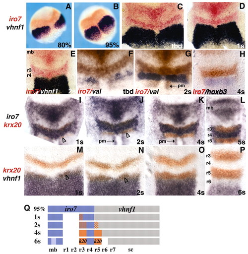
iro7 is expressed in a restricted domain of the neural plate with a posterior border at the prospective r4/r5 boundary. (A-P) Embryos stained by double in situ hybridisation with the indicated probes (colour coded). Anterior is towards the top, stages are indicated at the bottom right-hand corner of each picture. (A,B) Whole mounts; (C-P) flat mounts. The arrowheads in I,J,M,N indicate krx20-expressing cells in prospective r5. The arrows in G,J,K indicate a mesodermal domain of iro7 expression (pm for paraxial mesoderm). (Q) Summary of expression data, anterior is towards the left. mb, midbrain; sc, spinal cord. EXPRESSION / LABELING:
|
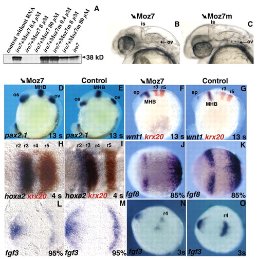
Blocking iro7 translation with Moz7 leads to a reduction of the midbrain and anterior hindbrain. (A) Autoradiography showing radiolabelled in vitro translation products of the iro7 capped RNA in the presence of increasing concentrations of Moz7 or Moz7m (0.4 μM to 80 μM). (B,C) Lateral view of live embryos at 30 hpf injected with Moz7 (B) or Moz7m (C), anterior is towards the left. (D-O) Embryos stained by in situ hybridisation with the indicated probes (colour coded). Anterior is towards the left, stages are indicated at the bottom right-hand corner of each picture. (B-G,J,K,N,O) Whole mounts; (H,I,L,M) flat-mounts. is, isthmus; ov, otic vesicle; os, optic stalk; MHB, midbrain-hindbrain boundary; ep, epiphysis. EXPRESSION / LABELING:
|

Knocking-down iro7 results in an anterior expansion of r5 at the expense of r4. (A-P) Whole-mount in situ hybridisation with the probes indicated (bottom left of each picture, colour coded) on embryos injected with Moz7 (A,C,E,G,I,K,M,O) or control embryos (B,D,F,H,J,L,N,P). Anterior is towards the left, except in E-H where anterior is towards the top. The inset in O presents a transverse section at the level of r4. The arrowheads indicate a group of cells expressing krx20 ectopically in the ventral part of r4. (C,D,K,L,M,N) Dorsal views of flat-mounted embryos. Stages are indicated at the bottom right of each picture. nc, neural crest. EXPRESSION / LABELING:
|
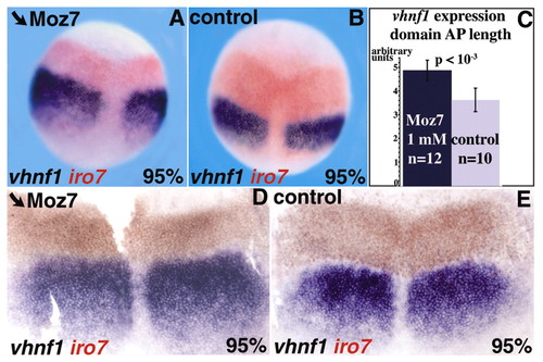
Knocking-down iro7 results in an anterior expansion of vhnf1 expression. (A,B,D,E) Whole-mount in situ hybridisation with the probes indicated (bottom left, colour coded) on Moz7 injected (A,D) or control (B,E) embryos. All pictures are dorsal views of whole-mount (A,B) or flat-mounted (D,E) embryos, with anterior towards the top. (C) A mean increase of 34% (t-test; P<0.001) of the AP length of vhnf1 expression domain in Moz7-injected embryos compared with control embryos. EXPRESSION / LABELING:
|
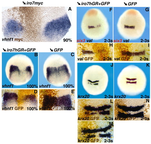
Ectopic expression of iro7 is sufficient to repress vhnf1, val and krx20. (A-N) Dorsal views of whole-mount (B,C,F,G,J,K) or flat-mounted (A,D,E,H,I,L,M,N) embryos analysed by in situ hybridisation and/or immunohistochemistry with the markers indicated (bottom left-hand corner of each picture, colour coded) on embryos injected with iro7myc (40 ng/μl) (A), iro7hGR (100 ng/μl) and GFP (B,D,F,H,J,L,M), or GFP alone (C,E,G,I,K,N). Anterior is towards the top. |

vhnf1 represses iro7 expression. (A-D) Double in situ hybridisation with probes for iro7 (blue) and krx20 (red) on vhnf1 homozygous (A,C) or control (B,D) embryos. (E) The statistical analysis of the posterior expansion of the iro7 expression domain in vhnf1 homozygous mutant embryos at 1-2 s. (F-H) In situ hybridisation with an iro7 probe on embryos injected with vhnf1 (20ng/μl) and lacZ (F,G) or with vhnf1Q139E (25 ng/μl) (H) RNAs. (G) Immunohistochemistry with an antibody directed against β-galactosidase. EXPRESSION / LABELING:
|

Knocking-down iro7 results in a reduction of neurogenesis in the anterior hindbrain. (A-D,G,H) Immunohistochemistry using anti-neurofilament RMO44 (A,B), 3A10 (C,D) or anti-Isl1 (G,H) antibodies on Moz7-injected (A,C,G) or control (B,D,H) embryos. (E,F) In situ hybridisation with a val probe on 20 hpf Moz7-injected (E) or control (F) embryos. (A-F) The r4-specific Mauthner neurons (‘M’, arrowhead in B,D,F) and the RoL2 neurons (arrows in B-D) are partially or totally lost after Moz7 injection (empty arrowheads and empty arrows in A,C,E). (I-L) Lateral views (I,J) or dorsal views (K,L) of live Isl1-GFP transgenic embryos injected with Moz7 (I,K) or uninjected (J,L). (G-L) nV, nVI, nVII and nX indicate the motor nuclei of the Vth, VIth, VIIth and Xth nerves, respectively. Arrows in I,J indicate the trigeminal (anterior) and facial (posterior) motor nerves. (M-P) Flat-mounted embryos analysed by in situ hybridisation with the probes indicated on the left (colour-coded) on Moz7-injected (M,O) or Moz7m-injected (N,P) embryos. Anterior is towards the left. Proneural clusters are indicated as follows: Mn, motoneurons; tg, trigeminal ganglion; RS, reticulospinal neurons; vcc, ventrocaudal cluster. EXPRESSION / LABELING:
|