
- Title
-
Fgf signalling is required for formation of cartilage in the head
- Authors
- Walshe, J. and Mason, I.
- Source
- Full text @ Dev. Biol.
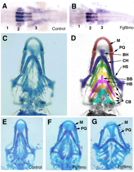
Effects of inhibition of Fgf8 on Hoxa2 expression and pharyngeal cartilage. (A, B) Hoxa2 expression in 18-hpf embryos following injection of control morpholino (A) or Fgf8mo (B). Transcripts are detected in the hindbrain and in the second (2) and third (3) neural crest streams that will populate the second (hyoid) pharyngeal arch and posterior arches, respectively, but are not detected in cells in the position of the first (1) stream. (C) Ventral view of a wild type embryo stained for cartilage at 5 days of development. (D) The same embryo as in (C) but with the skeletal derivatives of each arch differentiated by colour and individual elements labelled: arch 1, red; arch 2, blue; arch 3, green; arch 4, yellow; arch 5, pink; arch 6, orange; and arch 7, turquoise. (E–G) Ventral views of embryos injected with the control morpholino (E) or Fgf8mo (F, G) and allowed to develop to 5 dpf, then stained for cartilage. The eyes have been removed from (C, E–F) to improve visualization of the skeleton. Abreviations: M, Meckel's cartilage: PQ, palatoquadrate; BH, basihyal; CH, ceratohyal; HS, hyosymplectic; BB, basibranchial; HB, hypobranchial; CB, ceratobranchials. |
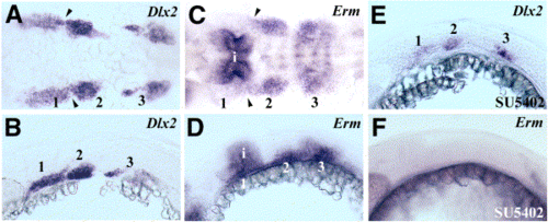
Migrating neural crest cells are recipients of Fgf signals. (A, B) Dorsal (A) and lateral (B) views of 24-hpf embryos showing expression of Dlx2 to show the locations of the three pharyngeal neural crest streams (numbered). (C, D) Dorsal (C) and lateral (D) views of 24-hpf embryos showing expression of Erm in the second and third neural crest streams but little expression in the first stream that migrates adjacent to the isthmus. Transcripts are also detected in the neural tube associated with the isthmus (i) and are detected more weakly in rhombomere boundaries in (C). (E, F) Treatment of embryos with 100 μM SU5402 between 18 and 24 hpf: Dlx2 expression shows that all three neural crest streams are still present (E) but Erm is undetectable (F). Arrowheads in (A) and (C) indicate the position of the first pharyngeal pouch. EXPRESSION / LABELING:
|
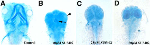
Fgf signalling is required for formation of pharyngeal and viscerocranial cartilages. (A–D) Ventral view of 4-dpf embryos stained to visualise cartilage following exposure to SU5402 for 24 h from the 12-somite stage (15 hpf). (A) Control embryo. (B–D) Embryos exposed to 10, 25, and 50 μM SU5402, respectively. A single, small cartilage element (arrow) is present in (B), and lenses are also present in that specimen (arrowhead) but these are absent from (C) and (D). |

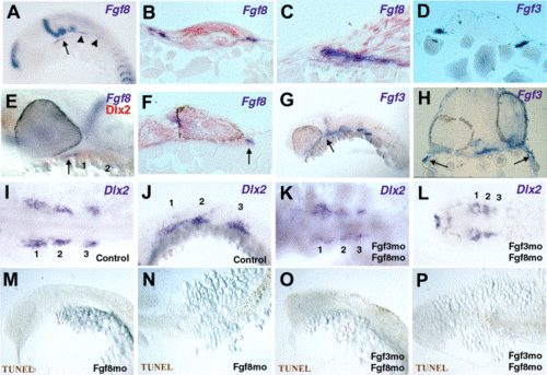
Fgf3 expression by pharyngeal pouch endoderm closely associated with neural crest streams. (A–H) Close relationship of endodermal Fgf3 expression to neural crest streams shown by hybridisation for Fgf3 (blue) and Dlx2 (orange) transcripts. Dorsal (A–D) and lateral (E–H) views of 18 (A, E), 21 (B, F), 24 (C, G), and 28 hpf (D, H) embryos. Fgf3 is expressed in endoderm of the first, second, and third pharyngeal pouches (arrows), also the isthmus (i) and rhombomere 4 (r4) in younger embryos (A, B, E, F) and in the anterior of the otic vesicle in older embryos (B–D, F–H; arrowhead in G, H). (I–L) Dlx2-positive neural crest cells are still present in embryos at 21 and 30 hpf following inhibition of Fgf3 translation with morpholinos. In situ hybridisation for Dlx2 (orange) and Fgf3 (blue) transcripts in embryos at 21 hpf injected with a control morpholino (I; dorsal view) or Fgf3mo (J; dorsal view). Arrows indicate first and third neural crest streams. Dlx2 expression at 30 hpf in embryos injected with a control morpholino (K; dorsal view) or Fgf3mo (L; dorsal view). (M–P) Posterior but not anterior expression of Erm by neural crest cells in 18-hpf embryos is reduced following inhibition of Fgf3. Dorsal (M, O) and lateral (N, P) views of embryos injected with control morpholino (M, N) or Fgf3mo (O, P). Erm transcripts are not detected in the most posterior of the neural crest streams (arrows). Isthmic Erm expression (i) is indicated in (M) and (O). (Q–T) Cell death is not detected in migrating crest streams at 20 hpf following inhibition of Fgf3. Dorsal (Q, S) and lateral (R, T) views of embryos injected with control (Q, R) or Fgf3mo oligonucleotides and analysed by TUNEL staining. No apoptotic cells are apparent in control embryos (Q, R), while death is restricted to dorsal neural tube at hindbrain and cord levels following inhibition of Fgf3 (S, T). EXPRESSION / LABELING:
|
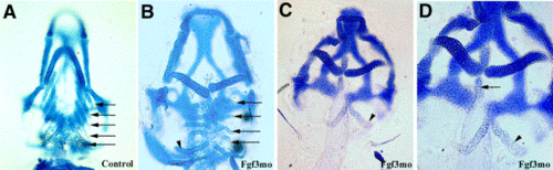
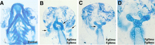
Fgf3 is required for formation of ceratobranchial cartilages. (A–D) Ventral views showing loss of cartilage derivatives of pharyngeal arches 3-6 in 5-dpf embryos following inhibition of Fgf3. (A) Embryo injected with control morpholino. (B) Embryo injected with low concentration of Fgf3mo shows reduction in ceratobranchial cartilages derived from arches 3-6 (arrows). (C, D) Low and high magnification images of an embryo injected with a higher concentration of Fgf3mo. There is a complete absence of all ceratobranchial cartilage derivatives of arches 3–6. Hypobranchial and basibranchial elements are also missing, although a small midline cartilage nodule is present (arrow in T). However, the tooth-bearing ceratobranchial element derived from arch 7 is unaffected (arrowheads in B–D). |
Endodermal expression of Fgf8 during early crest migration and coexpression with Fgf3 in endoderm associated with the jaw. (A) At 16 hpf, Fgf8 transcripts are detected in first pouch endoderm (arrow) and endoderm (arrowheads) associated with posterior arches. (B, C) Transverse sections through an embryo at 16 hpf showing Fgf8 expression in endoderm associated with posterior arches (B; region indicated by arrowheads in A) and with anterior arches (C; high power view of one side of a section from the region indicated by arrow in A). (D) Transverse section through the second pharyngeal pouch of a 21-hpf embryo shows Fgf3 expressed by pharyngeal endoderm. (E–H) At 30 hpf, both Fgf8 and Fgf3 are expressed by endoderm associated with the developing jaw. (E) In situ hybridisation for Fgf8 (blue) and Dlx2 (orange) transcripts at 30 hpf. Fgf8 (arrow) is expressed in cells immediately anterior to the first arch (arches 1 and 2 are indicated). (F) Oblique section through the forebrain, eye, and anterior arch region of an embryo at 30 hpf showing Fgf8 transcripts in endoderm (arrow). (G) Fgf3 is expressed in endoderm associated with anterior arches (arrow). (H). Transverse section through a 30-hpf embryo showing Fgf3 expression in anterior endoderm. (I–L) Dlx2-positive neural crest cells are still present in embryos at 21 and 30 hpf following inhibition of Fgf3 and Fgf8 translation with morpholinos. The three neural crest cell populations are numbered. In situ hybridisation for Dlx2 transcripts in embryos at 21 hpf injected with a control morpholino (I, dorsal view; J, lateral view). Three Dlx2-positive neural crest populations are detected at 21 hpf following inhibition of both Fgf3mo and Fgf8mo (K; dorsal view); however, at 30 hpf, expression is reduced in the posterior population (L; dorsal view). (M–P) Cell death is not detected in migrating crest streams at 20 hpf following inhibition of either Fgf8 alone (M, N) or both Fgf3 and Fgf8 (O, P). (M, O) Lateral views and (N, P) dorsal views. EXPRESSION / LABELING:
|
Fgf3 and Fgf8 are required together for formation of cartilage in the head. (A–D) Ventral views showing near-complete absence of pharyngeal and neurocranial cartilages in 5-dpf embryos in which both Fgf3 and Fgf8 are inhibited. (A) Ventral view of embryo injected with both control morpholinos. (B, C) Embryos in which both Fgf3 and Fgf8 have been inhibited show loss of most cartilage from the head; residual cartilage elements are indicated by arrows. (D) Higher magnification image of (C). |
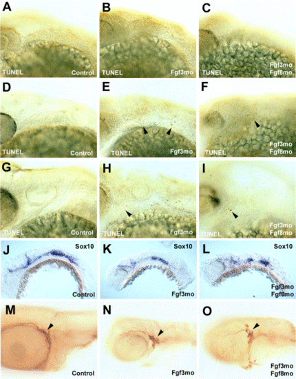
Effects of inhibition of Fgf3 or Fgf3 and Fgf8 on cranial neural crest cells. (A–I) Injection of Fgf3mo either singly or in combination with Fgf8mo causes little change in cell death in pharyngula stage embryos. Dead cells are extremely rare in the arch regions of embryos injected with control morpholinos at 25 (A), 30 (D), and 48 hpf (G). Injection of Fgf3mo or both Fgf3mo and Fgf8mo results in very little cell death in the arch region at 25 hpf (B, C), while more dead cells (arrowheads) are detected at 30 (E, F) and 48 hpf (H I). (J–O) Fgf3 and Fgf8 are not essential for nonectomesenchymal neural crest cells to express Sox10 or for formation of the trigeminal ganglion. (J–L) Neural crest cells which do not form cartilage express Sox10 in 24 hpf embryos injected with either control morpholinos (J), Fgf3mo (K), or both Fgf3mo and Fgf8mo (L). (M–O) Formation of the trigeminal ganglion (arrowheads) is unimpeded in embryos injected with either control morpholinos (M), Fgf3mo (N), or both Fgf3mo and Fgf8mo (O). |
Reprinted from Developmental Biology, 264(2), Walshe, J. and Mason, I., Fgf signalling is required for formation of cartilage in the head, 522-536, Copyright (2003) with permission from Elsevier. Full text @ Dev. Biol.