
- Title
-
Site-1 protease is required for cartilage development in zebrafish
- Authors
- Schlombs, K., Wagner, T., and Scheel, J.
- Source
- Full text @ Proc. Natl. Acad. Sci. USA

Cartilage defects in goz. Alcian blue staining of sibling larvae (a, c, and e) and goz larvae (b, d, and f) at 5 dpf is shown. (a and b) Lateral view. (c and d) Ventral view of the head. (e and f) Enlargement of the branchial arches. Cartilage matrix staining is slightly reduced in goz mutants. All cartilage elements, including Meckel′s cartilage (arrows in c and d), are smaller in the mutant. Tissue anterior to the eyes is missing (bars in c and d). The columnar arrangement of chondrocytes in the branchial arches is disrupted in goz mutants (arrows in e and f). (Scale bars are 200 µmin a-d and 50 µmin e and f.) PHENOTYPE:
|

Col II defects in goz. Immunohistological staining of Col II in wild-type (WT) sibling larvae (a, c, and e) and goz larvae (b, d, and f) is shown. (a and b) Ventral view of the head 5 dpf. (c and d) Enlargement of the trabeculae (tb) of the neurocranium (lateral view) at 2 dpf. (e and f) Enlargement of the notochord (n) in the tail at 2 dpf. Cartilage matrix is homogeneously stained in sibling larvae (a, c, and e), whereas abnormal protein aggregates can be seen in the cartilage matrix and around the notochord in goz larvae (d and f). (Scale bars are 200 µm in a and b and 50 µm in c-f.) PHENOTYPE:
|
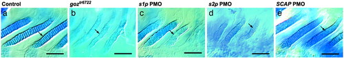
PMO phenocopy of goz phenotype. Shown are branchial arches of goz (b) or larvae injected with s1p (c) or s2p (d). PMOs show randomly arranged chondrocytes (arrows). Injection of SCAP PMOs resulted in wild-type cartilage (e), which is indistinguishable from injection controls (a). Larvae were stained with alcian blue at 5 dpf. (Scale bar is 50 µm.) PHENOTYPE:
|
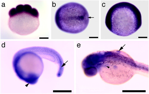
Expression pattern of S1P. Whole-mount in situ hybridization with s1p in eight-cell stage (a), tailbud stage (b), two-somite stage (c), 20 hpf (d), and 34 hpf (e) zebrafish embryos. (a, c, and d) Lateral view. (b) Dorsal view. (e) Dorsolateral view. Both maternal (a) and zygotic (b–e) expression of s1p can be observed. Expression is pronounced in the midline at tailbud stage (arrow in b). Strong expression can be seen in the head (d, arrowhead) and the immature notochord (d, arrow) at 20 hpf. At 34 hpf (e), s1p is strongly expressed in the head, the fin buds (arrow), and the region of the endodermal pouches (arrowhead). (Scale bar is 200 µm.) |
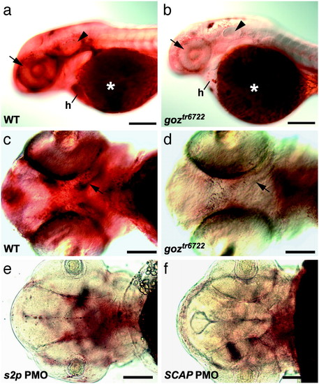
Lipid defects in zebrafish s1p mutants. Oil red O staining of 48 hpf zebrafish larvae. (a and b) Lateral view. (c-f) Ventral view. goz (b and d) and sibling (a and c) larvae and larvae injected with 6 ng of s2p2 (e) and 12 ng of SCAP1 (f) PMOs are shown. Strong lipid staining can be observed around the eye (arrow), the otic vesicle (arrowhead), and in the heart (h) of sibling larvae (a), in addition to the region of developing trabeculae (arrow in c). Both sibling and goz larvae show a strong lipid staining of the yolk (asterisks in a and b). Lipid deposit is severely reduced in all other tissues in goz larvae (b), especially around the eye (arrow) and the otic vesicle (arrowhead). No lipid can be detected around the developing trabeculae in goz (arrow in d). (e and f) Injection of s2p and SCAP PMOs results in the same reduction of lipid. (Scale bars are 200 µm in a and b and 100 µm in c-f.) PHENOTYPE:
|

Unillustrated author statements EXPRESSION / LABELING:
|