- Title
-
Radar is required for the establishment of vascular integrity in the zebrafish
- Authors
- Hall, C.J., Flores, M.V.C., Davidson, A.J., Crosier, K.E., and Crosier, P.S.
- Source
- Full text @ Dev. Biol.
|
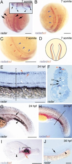
Expression of radar during embryogenesis and its relationship to scl expression. (A) Dorsal posterior view of radar expression in a seven-somite-stage embryo (anterior to top). (Inset) Posterior optical cross-section (posterior down) displaying mesodermal radar expression as bilateral stripes in the lateral plate (arrows) relative to more dorsal ectodermal expression above the neural keel (arrowheads). (B) Dorsal posterior view of a double in situ hybridization staining for radar (purple; purple arrowheads) and scl (red; red arrowheads) in a seven-somite-stage embryo. (C) A higher power view of embryo in (B). (D) Diagrammatic representation (dorsal posterior view) of radar and scl expression in the lateral mesoderm of a seven-somite-stage embryo. (E) Expression of radar in the trunk of a 24-hpf embryo (anterior to left) and its cross-section (F). The hypochord (white arrowhead) and posterior endoderm (white arrow) express radar, as well as the developing dorsal fin (red arrow) and dorsal neural tube (red arrowhead). (G) Expression of radar in the tail of a 24-hpf embryo was detected in the posterior hypochord (black arrow) and ventral tail mesenchyme (black arrowhead). (H) Double in situ hybridization staining for radar (purple) and scl (red) in the tail of a 24-hpf embryo and its cross-section (I) displays an overlap of expression in the ventral tail mesenchyme (bracket in I). (J) Lateral view of radar expression (anterior to left) in the trunk of a 36-hpf-stage embryo. Black arrowhead in (I) denotes notochord and the blue arrow, the hypochord. YE, yolk extension; N, notochord; NT, neural tube; DA, dorsal aorta; PCV, posterior cardinal vein. Scale bars in (A–C), (E, G, H, J), and (I) represent 150, 100, and 50 μm, respectively. EXPRESSION / LABELING:
|
|
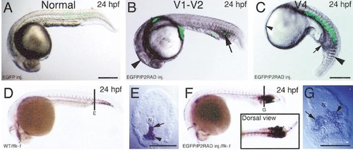
Forced radar expression elicits ventralized phenotypes. (A) Lateral view of a 24-hpf embryo following the injection of pCS2/EGFP (75-pg dose). The extent of mosaic EGFP expression is depicted as a superimposed green pattern in (A–C). (B) Lateral view of a mildly ventralized 24-hpf embryo (V1–V2) following the coinjection of P2RAD and pCS2/EGFP (75-pg dose). Low-level GFP expression is present in the posterior ICM region. Black arrowhead and arrow denote reduction of head structures and expanded ICM compartment, respectively. (C) Lateral view of a severely ventralized 24-hpf embryo (V4) following the coinjection of P2RAD and pCS2/EGFP (75-pg dose). Widespread GFP expression can be seen in the trunk. Small black arrowhead and arrow denote absent anterior head structures and expanded ICM region, respectively. Large black arrowhead denotes fused somites. (D) Whole-mount in situ hybridization staining for flk-1 in a 24-hpf wild type embryo and its cross-section through the developing tail (E). Arrow and arrowhead in (E) denote primitive caudal artery and caudal vein, respectively. (F) Expression of flk-1 in a mildly ventralized 24-hpf P2RAD/EGFP coinjected embryo (75-pg dose) and its transverse section through the tail (G). Arrows in (G) denote expanded flk-1 expression domain. N, notochord. Scale bars in (A, C) and (E, G) represent 200 and 100 μm, respectively. |
|
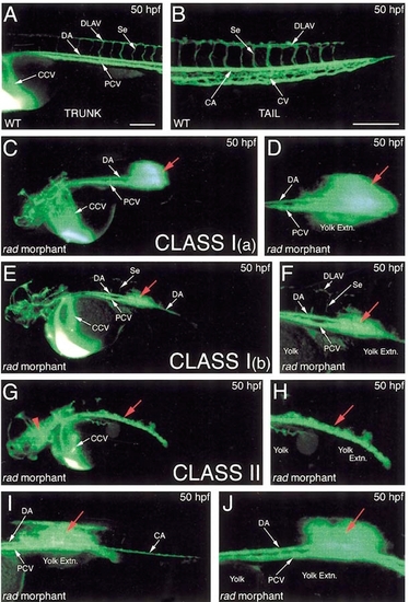
Visualization of Radar-specific circulation defects by microangiography. (A) Normal trunk circulation in a 50-hpf wild type embryo (anterior to left). (B) Normal caudal circulation in the tail of a 50-hpf wild type embryo (anterior to left). (C–J) Disrupted circulation observed in 50-hpf radar morphant embryos (injected with 8 ng rad-MO1). Red arrows denote hemorrhages. Red arrowhead denotes cranial hemorrhage. (D, F, H) The magnified views of embryos (C), (E), and (G), respectively. Embryos (C, E) and (G) represent morphants with class I and II circulatory defects, respectively. DLAV, dorsal longitudinal anastomotic vessel; DA, dorsal aorta; CCV, common cardinal vein; PCV, posterior cardinal vein; Se, intersegmental vessel; CA, caudal artery; CV, caudal vein. Scale bar represents 200 μm. PHENOTYPE:
|
|
The progression of vascular leaks from radar morphants that initiate normal circulation at 25 hpf. (A) Dorsal anterior view of a whole-mount in situ hybridization for βE3-globin expression in a 25-hpf radar morphant embryo. Large black arrows and arrowheads in (A) denote blood cells in the common cardinal vein and the anterior extremities of the posterior cardinal vein, respectively. Small black arrows in (A) denote blood cells in the left and right lateral dorsal aorta. (B) Microangiography displaying normal posterior circulation in a 25-hpf radar morphant embryo. (C) Lateral view of a 35-hpf radar morphant embryo (anterior to left) displaying the genesis of a vascular leak. Red arrows denote direction of blood flow. (D) A higher power view of embryo in (C). Red arrowheads denote blood cells just dorsal to the notochord that have leaked from the dorsal aorta. (E) Cross-section of a 50-hpf wild type embryo (dorsal to top). (F) Magnified dorsal view of cross section in (E). (G) Similar view of a cross-section from a 50-hpf radar morphant, displaying red blood cells pooling around the dorsal extremities of the trunk (black arrowheads). DA, dorsal aorta; PCV, posterior cardinal vein; CA, caudal artery; CV, caudal vein; NT, neural tube; N, notochord; RBC, red blood cell. Scale bars in (A, B), (C), (E), and (F, G) represent 200, 100, 50, and 20 μm, respectively. |
|
Normal vascular patterning and hematopoiesis in radar morphant embryos. Whole-mount in situ hybridization analysis of flk-1 (A, B), ang1 (C, D), fli1 (E, F), tie2 (G, H), and βE3-globin (I, J) in wild type (A, C, E, G, I) and radar morphant (B, D, F, H, J; injected with 8 ng rad-MO1) embryos. DA, dorsal aorta; PCV, posterior cardinal vein; Se, intersegmental vessel. Scale bars in (A) and (I) represent 250 and 200 μm, respectively. |
Reprinted from Developmental Biology, 251(1), Hall, C.J., Flores, M.V.C., Davidson, A.J., Crosier, K.E., and Crosier, P.S., Radar is required for the establishment of vascular integrity in the zebrafish, 105-117, Copyright (2002) with permission from Elsevier. Full text @ Dev. Biol.


