
- Title
-
Eye defects in receptor protein-tyrosine phosphatase alpha knock-down zebrafish
- Authors
- van der Sar, A.M., Zivkovic, D., and den Hertog, J.
- Source
- Full text @ Dev. Dyn.
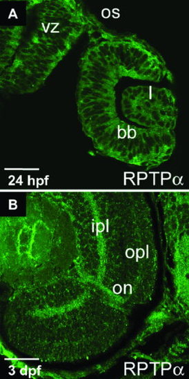
Expression of receptor protein-tyrosine phosphatase alpha (RPTPα) in the zebrafish eye. Indirect immunofluorescence, by using affinity purified anti-RPTPα antibody (AP5478) of a 24 hours postfertilization (hpf) embryo (A) and a 3 days postfertilization (dpf) embryo (B). Close-up photomicrographs of the eye are shown. bb, basal border; ipl, inner plexiform layer; on, optic nerve; opl, outer plexiform layer; os, optic stalk; l, lens; vz, ventricular zone. Scale bars = 150 μm in A, 40 μm in B. EXPRESSION / LABELING:
|
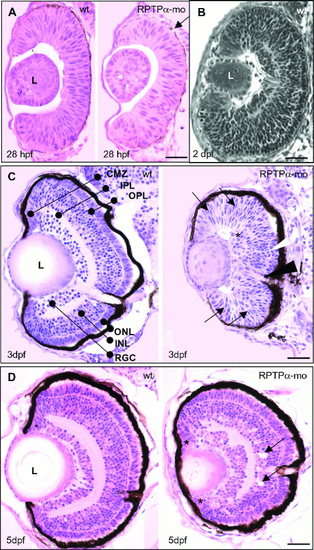
Receptor protein-tyrosine phosphatase alpha (RPTPα) knock-down–induced defects in eye development. Retinal organization of wild-type (left panels) and RPTPα-morpholino (-mo) -injected embryos (right panels). Transverse median sections through the eye of embryos at different developmental stages. A: At 28 hours postfertilization (hpf), the neural retina of RPTPα-mo–injected embryos consists of fewer cells compared with wild-type (wt) retinae. The arrow indicates the pigmented neural epithelium. B: Transverse section of a 2-days postfertilization (dpf) -old embryo. The retina already shows lamellar organization (see also: http://imaging.niob.knaw.nl/). C: At 3 dpf, the neural retina of RPTPα-mo–injected embryos are largely undifferentiated (arrows) and only in the central region some rudimentary lamellar organization can be observed. Overall, the size of the eye is reduced compared with wild-type embryos. The asterisk indicates potential retinal ganglion cells, the white arrowhead indicates potential outer plexiform layer, and the black arrowhead indicates the optic nerve. D: At 5 dpf, the retinae of RPTPα-mo–injected embryos contain five layers and most cells are differentiated. Asterisks indicate the ciliary marginal zones, arrows indicate gaps in the amacrine cell layer of the inner nuclear cell layer. At least three embryos were examined per stage, and representative sections are depicted here. L, lens; CMZ, ciliary marginal zone; RGC, retinal ganglion cells; IPL, inner plexiform layer; INL inner nuclear layer; OPL, outer plexiform layer; ONL, outer nuclear layer. Scale bars = 50 μm in A,C,D, 60 μm in B. |
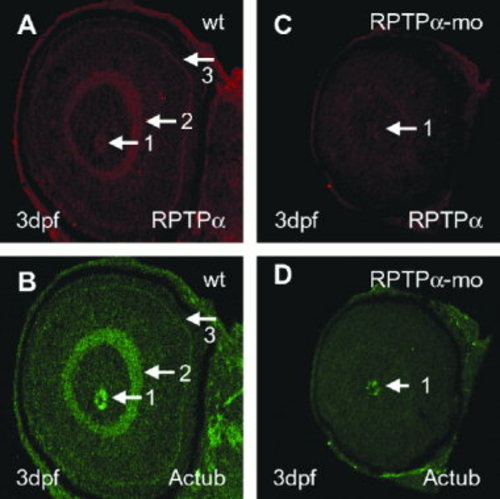
Defects in retinal organization and differentiation in receptor protein-tyrosine phosphatase alpha morpholino (RPTPα-mo) -injected embryos. A,B: Oblique section of 3-days-postfertilization (dpf) -old wild-type (wt) embryo labeled with polyclonal anti-RPTPα antibody (AP5478; A) and anti-acetylated tubulin (B). Labeling is found in the optic nerve (arrow 1), the inner plexiform layer (arrow 2), and the outer plexiform layer (arrow 3). C,D: Oblique section of 3-dpf-old RPTPα-mo–injected embryo labeled with polyclonal anti-RPTPα antibody (AP5478; C), and anti-acetylated tubulin (Actub) antibody (D). Labeling of RPTPα and acetylated tubulin is found in the optic nerve (arrow 1), all other structures labeled in wild-type are not labeled in RPTPα-mo–injected embryos. EXPRESSION / LABELING:
PHENOTYPE:
|