
- Title
-
Expression of the zebrafish genome during embryogenesis (NIH R01 RR15402)
- Authors
- Thisse, B., Pflumio, S., Fürthauer, M., Loppin, B., Heyer, V., Degrave, A., Woehl, R., Lux, A., Steffan, T., Charbonnier, X.Q. and Thisse, C.
- Source
- Submitted By
- Obrecht-Pflumio, Sophie, Thisse, Bernard, Thisse, Christine (Citing this work)
- Protocol
- Thisse in situ hybridization protocol
- Probe
- cb643 Quality:
- Supplier
-
Supplier: Zebrafish International Resource Center (ZIRC) (order this)


Fig. 1 snail1 is maternally expressed and maternal transcript are observed all along blastula stage. Zygotic expression of snail1 appears at the dome stage first in the dorsal most marginal blastomere then the expression extend to encompass all the blastula margin EXPRESSION / LABELING:
|


Fig. 2 Expression of snail1 is observed at the gastrula margin as well as in presumptive paraxial mesoderm. Snail1 transcript are excluded from the dorsal most territories (central part of the embryonic shield, axis) EXPRESSION / LABELING:
|


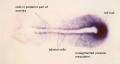
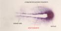
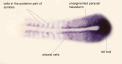
Fig. 3 Expression of snail1 is specific of segmental plate and tail bud. Snail is first strongly expressed in adaxial cells, posterior unsegmented paraxial mesoderm and tail bud. When somites start to form, expression appears in the posterior cells of somites which line the somitic furrow. EXPRESSION / LABELING:
|


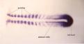

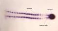


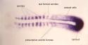


Fig. 4 Expression of snail1 is observed in somites, unsegmented paraxial mesoderm and tail bud. Expression of snail1 disappears from adaxial cells when somites form. and A second territory of snail1 expression appears in the head at the level of head mesoderm that give rise to head mesenchyme. In the unsegmented paraxial mesoderm, snail1 appears more strongly expressed in cells at the level of the presumptive somitic furrow (for the two presumptive somites posterior to the last somitic furrow formed) EXPRESSION / LABELING:
|


Fig. 5 Expression of snail1 decreases in myotomes as muscle cells differentiates. Snail1 start to be strongly expressed in head mesenchyme and all branchial arches EXPRESSION / LABELING:
|


Fig. 6 Expression of snail1 is strong in branchial arches and in pectoral fin bud. |


Fig. 7 In pectoral fin, snail1 is expressed in the mesenchyme under the apical ectodermal ridge as well as in the pectoral fin muscles. Snail 1 expression is also observed in the pharynx, oesophagus and intestinal bulb. |