
- Title
-
dackel acts in the ectoderm of the zebrafish pectoral fin bud to maintain AER signaling
- Authors
- Grandel, H., Draper, B.W., and Schulte-Merker, S.
- Source
- Full text @ Development

Phenotype of dak and syu mutants at 72 h (A,B), 38 h (C,D) and 96 h (E-H): (A,C,E) Wild-type (wt), (B,D,F) dak-, (G) syutq252, (H) syut4. (A,B) anterior is towards the left, (C,D) anterior is towards the left, view from distal. Note that the wt apical fold in C produces a ‘line’ (arrow) where dorsal and ventral epidermis become juxtaposed. This ‘line’ is absent in the dak- bud in D. (E-H) distal is towards the right. Living dak- larvae lack functional pectoral fins at 72 h and fail to develop an apical fold at 38 h. At 96 h dak- larvae have developed a pectoral girdle that distinguishes them from syu hypomorphic and null mutants. cl, cleithrum; ed, endoskeletal disc; ff, fin fold; pg, shoulder girdle; pp, postcoracoid process. |

Expression of apical ectodermal markers at 28 h (A,B,E) and 38 h (C,D,F,G,H,I,K-N). Anterior is towards the left, distal is towards the top. (J) shows the expression at 49 h: distal is towards the right, dorsal is towards the top. (A,C,F,H,J,K,M) wt, (B,D,G,I,L) dak-, (E) wt and dak- are indistinguishable, (N) syut4. (A-D) expression of dlx2. (E-G) expression of bmp2. (H-J,M,N) expression of fgf4. (K,L) expression of fgf8. dlx2 and bmp2 are activated at 28 h but their expression is strongly reduced at 38 h in dak-. Both fgfs are not properly activated in dak-. fgf4 is not expressed in syut4. |

Expression of syu and ptc1 at 28 h (A,B,G,H), 38 h (C,D,I,J) and 48 h (E,F,K,L). (A,C,E,G,I,K) wt and (B,D,F,H,J,L) dak- fin buds. Anterior is towards the left, distal is towards the top. The expression of both genes is reduced at 28 h and 38 h. No expression is detected at 48 h. |
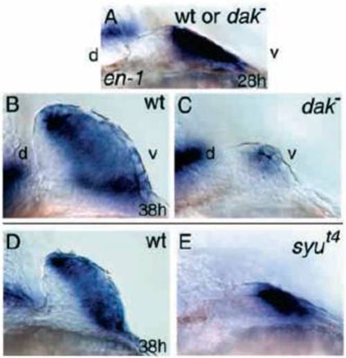
en1 expression in dak- and syut4 at 28 h (A) and 38 h (B-E). (B,D) wt, (C) dak- and (E) syut4. (A) wt and dak- are indistinguishable. Dorsal is to the left and ventral is to the right. At 28 h, dak- shows wt levels of en1. At 38 h, expression of en1 is downregulated in dak- but not in syut4. |

Expression of hoxd genes at 28 h (A,F,G,L,M), 32 h (R,S), 38 h (B,C,H,I,N,O,T,U) and 48 h (D,E,J,K,P,Q,V,W). Anterior is towards the left, distal is towards the top. (B,D,F,H,J,L,N,P,R,T,V) wt and (C,E,G,I,K,M,O,Q,S,U,W) dak-. (A) dak- and wt are indistinguishable. (A-E) expression of hoxd10. (F-K) expression of hoxd11. (L-Q) expression of hoxd12. (R-W) expression of hoxd13. Hoxd gene expression is lost at 48 h in all cases. Note the downregulation of hoxd10 at 38 h which is not seen in syut4. |
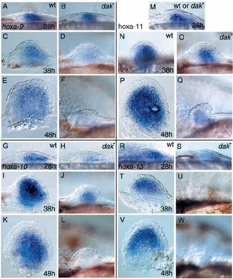
Expression of hoxa genes at 28 h (A,B,G,H,M,R,S), 38 h (C,D,I,J,N,O,T,U) and 48 h (E,F,K,L,P,Q,V,W). Anterior is towards the left, distal is towards the top. (A,C,E,G,I,K,N,P,R,T,V) wt and (B,D,F,H,J,L,O,Q,S,U,W) dak-. (M) dak- and wt are indistinguishable. (A-F) expression of hoxa9. (G-L) expression of hoxa-10. (M-Q) expression of hoxa-11. (R-W) expression of hoxa13. hoxa9 and hoxa10 are lost at 48 h, while slight hoxa11 and hoxa13 expression may still be detectable |
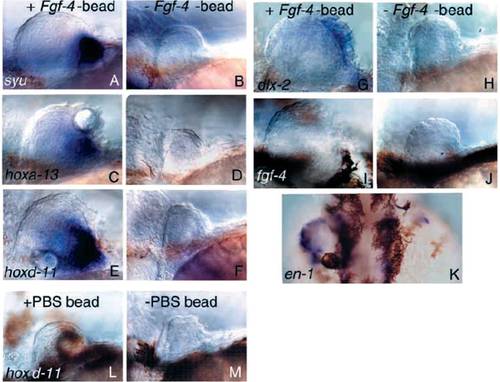
Implantation of an Fgf4-soaked bead (A,C,E,G,I,K) or a PBS-soaked control bead (L) into dak- fin buds. (B,D,F,H,J,M) Contralateral fin bud of the same embryo not treated with Ffg4- or PBS-bead. Marker gene expression is shown at 60 h. (A,B) syu, (C,D) hoxa13, (E,F,L,M) hoxd11, (G,H) dlx2, (I,J) fgf4. Anterior is towards the left and distal is towards the top. (K) en1. Distal view, anterior is towards the top. All Fgf4 bead-treated buds are larger than the untreated buds on the contralateral side. All mesenchymal markers tested are activated. In the apical ectoderm only early markers are activated (dlx2, bmp2; see text) whereas late markers are not activated (fgf4, fgf8; see text). Note that en1 is activated. PBS-bead implantation does not lead to marker gene activation of control buds. |
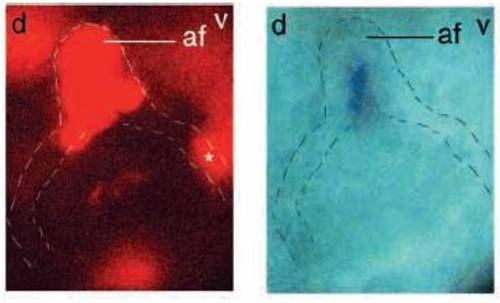
Transplantation of wt cell into a dak- fin bud. Distal is towards the top, dorsal towards the left and ventral towards the right. Fluorescent image (left) shows biotin-labeled wt cell in the apical fold (af) and in the ventral non-ridge ectoderm (*). Bright-field image (right) shows expression of fgf8 in the apical fold. The outline of the bud and the epidermal basal lamina are indicated by the broken lines. |

Unillustrated author statements PHENOTYPE:
|