
- Title
-
Multiple pathways in the midline regulate concordant brain, heart and gut left-right asymmetry
- Authors
- Bisgrove, B.W., Essner, J.J., Yost, H.J.
- Source
- Full text @ Development

Two-color double in situ hybridization identifies overlapping asymmetric gene expression patterns in the heart field (A-D) and diencephalon (E-J) of 22- to 24- somite stage zebrafish embryos. Ventral views, anterior at top, of lft2 expression (in red) and nkx2.5 (A), bmp4 (B), cyc (C) or pitx2 (D) expression (in purple), in the heart field. (E-J) Coexpression of lft1 (in red) with cyc (E-G, in purple) and pitx2 (H-J, in purple) in the left diencephalon (E, H, lateral views, dorsal at right; F, I, dorsal views, anterior at top; G, J, transverse sections, dorsal at top). EXPRESSION / LABELING:
|

Examples of altered patterns of asymmetric lft1 and lft2 expression in zebrafish midline mutant embryos. In situ hybridization of lft1 and lft2 in the diencephalon and heart field of 22- to 24-somite stage wild-type and mutant zebrafish embryos. Embryos, shown in dorsal view, anterior at top, have been removed from the yolk cell. (A) Wild-type, (B, C) ntlb160, (D) mom, (E) din, (F) cyctf219, (G) cycb229, (H) oep. EXPRESSION / LABELING:
|
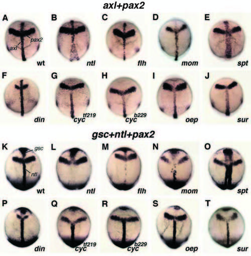
Expression patterns of midline mesendodermal marker genes are altered in zebrafish midline mutant embryos. Dorsal views, anterior at top, of tailbud-stage embryos, probed with axl (A-J) and gsc + ntl (K-T). All embryos were also probed with pax2, which is expressed in stripes that extend laterally from the midline, as a reference of anteroposterior position. (A,K) wild-type. (B-D, L-N) Class I: (B,L) ntlb160, (C,M) flh, (D, N) mom. (E,F,O,P) Class II: (E,O) spt, (F,P) din. (G,H,Q,R) Class III: (G,Q) cyctf219, (H,R) cycb229. (I,J,S,T) Class IV: (I,S) oep and (J,T) sur. EXPRESSION / LABELING:
|