
- Title
-
Zebrafish no isthmus reveals a role for pax2.1 in tubule differentiation and patterning events in the pronephric primordia
- Authors
- Majumdar, A., Lun, K., Brand, M., and Drummond, I.A.
- Source
- Full text @ Development
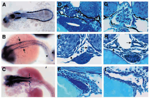
Fig. 1. noi affects multiple aspects of pronephric development. (A-C) Pattern of pax2.1 expression in the pronephros during wild-type development. Whole-mount in situ hybridization using the pax2.1 antisense probe on (A) 12 somite, (B) 24 hpf and (C) 30 hpf wildtype embryos. pax2.1 is first expressed in intermediate mesoderm (A), and then pax2.1 expression is maintained in anterior duct (black arrows in B) and forming cloaca (arrowhead in B). Later, pax2.1 is transiently expressed in forming pronephric tubules (arrows in C). Transverse histological sections of (D-F) wildtype and (G-I) noi- mutant embryos at (E,H) 36 hpf and (D,F,G,I) 3 dpf. In wild type, tubules extend laterally from a medial glomerulus and pronephric duct (D). In noi- mutants, pronephric tubules are missing even though a midline glomerulus is seen (G). Pronephric duct is not seen at this level of section. Wild-type pronephric ducts (arrows in E) have well-formed epithelial cells and a small central lumen. In noi-, the pronephric ducts (arrows) are distended and epithelia appear flattened (H). Sagittal histological section of wildtype pronephric cloaca shows cuboidal epithelial cells and a central lumen (F). Cloaca morphogenesis in noitu29 is affected (I). Epithelial cells are flattened and the lumen is greatly distended. Note the lumen does open to the outside. EXPRESSION / LABELING:
PHENOTYPE:
|
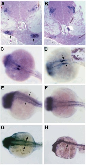
Fig. 4. noi affects patterning of the nephric primordia. (A,C,E,G) Wildtype and (B,D,F,H) noitb21 embryos in situ hybridized with pax2.1 (A,B,G,H), wt1 (C,D) and a-subunit of Na+/K+ ATPase antisense probes (E,F) at 30 hpf (A,B,G,H) or 25 hpf (C-F). (A) Transverse histological sections (schematized by A in Fig. 3) of in situ hybridized 30 hpf embryos shows expression of pax2.1 in lateral, but not medial, cells of the pronephric primordia in wild type. (B) pax2.1 expression in nephric primordia is greatly reduced in noitb21 mutants, even though CNS expression still remains. (C) Whole-mount in situ hybridization with wt1 marks the nephric primordia in wild type. wt1 expression is caudally expanded in noitb21 (arrows in D). Sagittal section through a noitb21 pronephros stained with wt1 antisense probe (inset in D) reveals ectopic wt1 expression in lateral cells of the nephron primordia (arrows). Anterior duct expression of a-subunit of Na+/K+ ATPase (arrows in E) is lost in noitb21 embryos while more posterior duct expression remains (F). Whole-mount in situ hybridization with pax2.1 reveals tubule and duct expression in wild type (arrows in G). (H) Tubule expression is gone, but anterior duct pax2.1 expression remains in noitb21. EXPRESSION / LABELING:
|
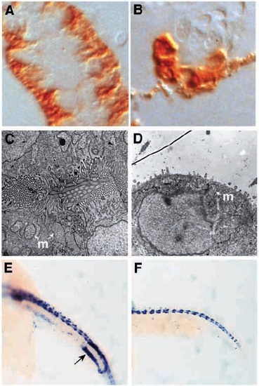
Fig. 6. noi mutants show defects in pronephric duct epithelial differentiation and cloaca development. (A,C,E) Wild-type and (B,D,F) noi- embryos stained with a-6F mAb at 3 dpf (A,B) and electron micrographs of pronephric duct epithelia from 2 dpf embryos (C,D). (A) In wild-type pronephric tubule and duct, mAb a- 6F staining is basolateral reflecting the basolateral subcellular localization of Na+/K+ ATPase a-subunit. (B) In noi mutants, mAb a-6F staining is diffuse throughout the duct cells with mAb a-6F positive cells sitting adjacent to duct cells showing no mAb a-6F reactivity. Some endogenous pigmentation is seen in B and is not HRP reaction product. Electron micrograph of wild-type pronephric duct epithelia shows an abundance of microvilli and cilia packed densely in the lumen. Mitochondria (m) are excluded from the apical terminal web. In noi-, the pronephric duct lumen is greatly distended and epithelia lack a apical brush border seen in D as a huge reduction in apical microvilli. Some ciliated cells are still present. Apically localized mitochrondria (m) are seen. Cloaca development is abnormal in noi mutants. (E,F) In situ hybridization with ret1 antisense probe at 22 hpf. ret1 is expressed in a posterior domain of the pronephric duct during cloaca development (arrow in E). Ductspecific expression of ret1 is absent in noi– even though CNS expression remains unaffected (F). EXPRESSION / LABELING:
PHENOTYPE:
|