
- Title
-
Equivalent genetic roles for bmp7/snailhouse and bmp2b/swirl in dorsoventral pattern formation
- Authors
- Schmid, B., Fürthauer, M., Connors, S.A., Trout, J., Thisse, B., Thisse, C., and Mullins, M.C.
- Source
- Full text @ Development
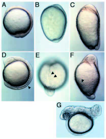
Morphological features of the snh mutant. Bud-stage embryos of (A) wild type, (B) snhty68a and (C) snhsb1aub mutants. 7-somite stage (D) wild-type embryo; (E) snhty68a mutant embryo (dorsal, posterior view), where the first two somites are laterally enlarged, but do not reach ventral regions, while more posterior somites do. (F) snhsb1aub mutant (oblique view), where all somites extend to ventral positions. Arrowheads mark the somites in D-F. (G) Same snhty68a embryo as in E at 1 d.p.f. (A-D) are lateral views, anterior to the top. PHENOTYPE:
|

The bmp7 gene in snhsb1aub, Df(LG11)snhp11 and Df(LG11)snhp15 alleles. (A) DNA of wild-type (+/+), heterozygous (+/-), and homozygous (-/-)snhsb1aub genotypes was digested and analyzed by genomic Southern blot, probed with the bmp7 3′UTR. This shows that the snhsb1aub chromosome is associated with an alteration of the bmp7 locus. (B) No bmp7 3′UTR PCR amplification product is observed in homozygous Df(LG11)snhp11 (Dfp11) and Df(LG11)snhp15 (Dfp15) mutant embryos, while it is amplified from wild-type sibling DNA. The bottom panels show an amplification product for the tolloid (tld) gene, confirming the integrity of the DNA. (C) The markers Z8214, Z13411 and Z6909 were absent in Df(LG11)snhp11 and Df(LG11)snhp15 homozygous mutants (lanes 1-3 of each gel), while they were present in wild-type siblings (lanes 4-6). Marker Z13395 was present in DNA of mutant and wild-type siblings. |

bmp7 expression during embryogenesis. (A) 30% epiboly stage (lateral view). Expression extends throughout the blastoderm, except its dorsal marginal aspect (arrowhead). Animal pole (B) and lateral (C) views of a germ ring stage embryo showing bmp7 downregulation in the dorsal quadrant. (D-F) 60% epiboly stage. Lateral (D) and transverse optical cross-sectional (E) views showing expression throughout the ventral gastrula and in the prechordal plate (pp) (D). (F) Close-up of the ventral margin showing expression in the YSL. (G) Lateral view at 90% epiboly. Robust expression is seen at the border between epidermal (epi) and neurogenic (neur) ectoderm and in the ventral margin. Faint expression is observed throughout the epidermal territory. (H,I) 3-somite stage. (H) Dorsal cephalic view. Transcripts accumulate at the border of the hatching gland (hg). Faint expression is detected outside the neural plate. (I) Posterior view. Transcripts are localized to the tail bud (tb) and the border between neural and non-neural posterior ectoderm. At 24 h.p.f. expression is observed in (J) the endoderm (end) and the pineal gland (p), (K) the posterior aspect of the otic vesicle (ov) and (L) dorsal posterior neural tissue (spc) of the tail. A-G, dorsal to the right; H-K, anterior to the left. EXPRESSION / LABELING:
|
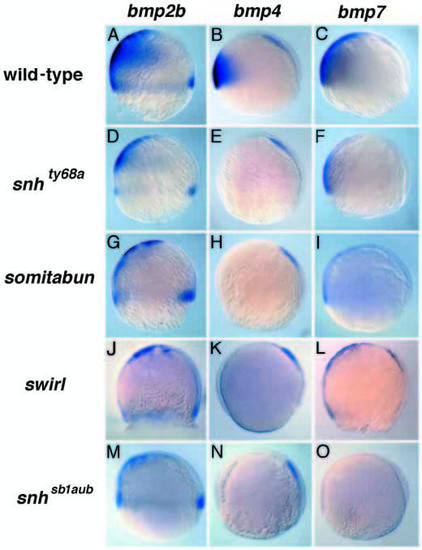
Expression of bmp2, 4 and 7 in dorsalized mutant embryos at 65-75% epiboly. Expression in wild-type embryos of bmp2b (A), bmp4 (B), and bmp7 (C). Expression of bmp2b, bmp4 and bmp7 in mutant embryos of snhty68a (D-F), sbndtc24 (G-I), swrta72/bmp 2b (JL) and snhsb1aub (M-O), respectively. In snhty68a mutants bmp2b (D) and bmp7 (F) expression are reduced in amount and to a more ventral domain. bmp4 expression is absent in snhty68a(E), sbn (H), swr (K) and snhsb1aub (N) on the ventral side, whereas the dorsal expression domain is unaffected. bmp2b expression is absent in the hypoblast ventrally, but is still present in the YSL and the dorsal margin in sbn/smad5 (G), swr/bmp2b (J) and snhsb1aub (M) mutant embryos. bmp7 expression is reduced in sbn/smad5 (I) and absent in swr/bmp2b mutant embryos, except for expression in the YSL and prechordal plate (L). In snhsb1aub mutants there is no detectable bmp7 expression, including the YSL and prechordal plate expression domains (O). EXPRESSION / LABELING:
|
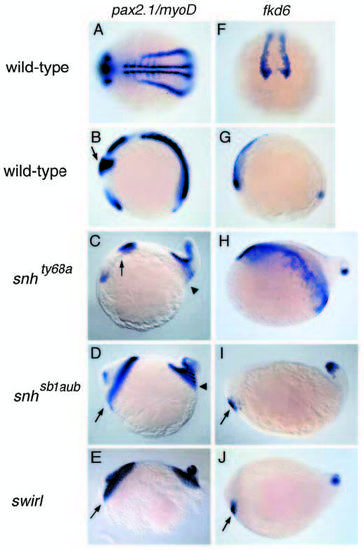
In situ hybridizations with pax2.1 and myoD (A-E) and fkd6 (F-J) in wild-type, snh/bmp7 and swrta72/bmp2b mutant embryos at the 5-somite stage. Lateral views, anterior to the left, except (A,F). (A) The somitic expression of myoD and the pronephric expression of pax2.1 in a dorsal-posterior view of a wild-type embryo. (B) The midbrain/hindbrain boundary (MHB) expression of pax2.1 (arrow) in a wild-type embryo. (C) snhty68a displays a slight lateral expansion of the MHB (arrow), not visible in this lateral view. The somites posterior to somite 2 encircle the embryo (arrowhead). (D) In snhsb1aub mutant embryos the MHB (arrow) and all somites (arrowhead) encircle the embryo. (E) Homozygous swrta72 mutants are indistinguishable from snhsb1aub mutants. fkd6 expression in the cranial neural crest progenitors in wild-type in an anterior (F) (dorsal to the top) and lateral (G) view. fkd6 expression is also observed in the tail bud (G). (H) Expanded cranial neural crest expression in snhty68a. In snhsb1aub (I) and swr/bmp2b (J) mutants, the number of cranial neural crest progenitors is severely reduced (arrow). EXPRESSION / LABELING:
|
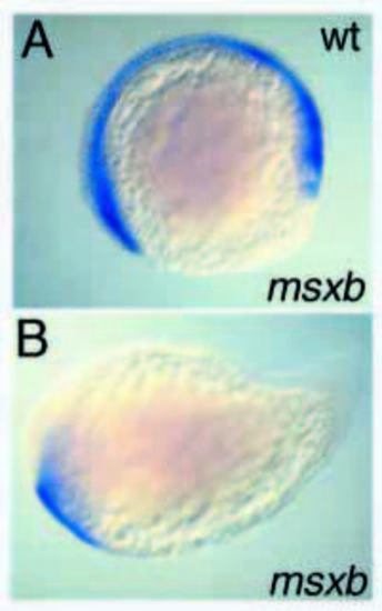
Expression of msxB in swr/bmp2b; snh/bmp7 double mutant embryos. Expression of msxB in 5- somite stage (A) wild-type and (B) mutant embryos of crosses between swrtc300a/+; snhsb1aub/+ double heterozygous fish. EXPRESSION / LABELING:
|

Bmp2b and Bmp7 interact synergistically in a cellautonomous manner. (A-D) Lateral views of live 24 h.p.f. embryos of the four phenotypic classes resulting from injection of bmp mRNAs. (A) Class I embryos display wild-type morphology. (B) In class II embryos the head is slightly reduced, while the hematopoietic mesoderm is expanded (arrow). (C) Class III embryos lack head and notochord. (D) Class IV embryos acquire a spindle shape lacking obvious dorsoventral polarity. (E) Phenotypic distributions following the expression of different bmps either separately or in combination. A synergistic increase in ventralizing potential is observed following the coexpression of bmp2b or bmp4 with bmp7, but not following the coexpression of bmp2b with bmp4. (F) Separate injection of one dose of bmp2b RNA and one dose of bmp7 RNA in adjacent blastomeres (lower, left panel) does not cause an enhanced effect compared to the injection of two doses of bmp2b or bmp7 (upper panels). A cooperative effect between bmp2b and bmp7 is only observed if RNAs for both factors are coinjected in the same cell (lower, right panel). |